
- 1. The lumps vets actually see
- 2. Abscess and infected lump
- 3. Cysts, lipomas and warts
- 4. Tumours, and the ones that must come out
- 5. How a vet tells them apart
- 6. Mammary lumps in older female dogs
- 7. After removal: the cavity and the skin
- 8. Clinical cases
- Frequently asked questions
- Related guides
1. The lumps vets actually see
Most lumps on dogs fall into a small number of categories, and they differ in how fast they arrive, how they feel and how urgent they are:
| Lump | How it presents | Usual approach |
|---|---|---|
| Abscess | Appears over one to three days, warm and painful to touch, soft or fluid-filled in the middle, often with a recent bite, puncture or foreign body | Drained and flushed by a vet, left open to heal from the inside. Full guide → |
| Cyst (sebaceous or follicular) | Round, smooth, movable, slow-growing, not painful; sometimes discharges a grey-white paste | Often monitored; removed if it keeps filling, becomes infected or annoys the dog |
| Lipoma | Soft, movable, lobulated, under the skin, most common in middle-aged and older dogs | Usually left alone once confirmed. Removal only if it is large, awkward or growing quickly |
| Histiocytoma | A rapidly appearing red, button-like nodule in a young dog, often on the head or legs | Usually regresses on its own within one to three months; confirmed by needle aspirate rather than removed in a hurry |
| Mast cell tumour | Highly variable — can look like anything, may swell and shrink from day to day, often on the trunk or limbs | Surgical removal with wide margins. One of the diagnoses that must not be watched |
| Mammary tumour | A firm nodule in the mammary chain of a middle-aged or older female dog | Removed. Roughly half are malignant, and early removal is the single biggest factor in outcome |
| Warts and papillomas | Cauliflower-like, often multiple, common in young dogs | Usually regress as immunity develops; removed only if they bleed or interfere |
| Perianal adenoma | A lump beside the anus, usually in an entire male dog | Removed, and castration is part of the plan in most cases |
The point of the table is not to let you match your dog’s lump to a row. It is to show that the appearance and the feel of a lump overlap so heavily between harmless and serious that no owner — and no veterinarian — can identify a lump by touching it. The vet’s judgement comes from a needle and a microscope, not from the fingers.
2. Abscess and infected lump
An abscess is a walled-off pocket of pus, and it behaves differently from almost everything else on the list: it arrives fast, and it hurts.
- Timing. One to three days from nothing to an obvious swelling — often the owner can say which day they first noticed it.
- Feel. Warm, painful, and soft or fluid-filled in the centre, with firmer walls. The dog may resent being touched, and may be quiet or off its food.
- Cause. Usually a bite or puncture wound that sealed over, a foreign body such as a grass seed, an infected tooth root, or an infected anal sac. The infection was introduced days before the lump appeared.
- What happens to it. It is drained, the cavity is flushed, and it is deliberately left open to heal from the inside out. The one thing that must not be done is stitching it shut again, which reseals the infection and recreates the abscess.
Do not squeeze or lance it at home. Pressure drives the infection deeper into the tissue planes rather than out, it is very painful, and it risks seeding infection into a larger area. And if the swelling is not an abscess but a tumour or a cyst, squeezing is worse than useless. Wait for a diagnosis.
The overlap with tumours is real in both directions. An abscess that has been present for two weeks and is no longer hot or painful can be mistaken for a tumour; a tumour that has grown quickly and become inflamed can be mistaken for an abscess. This is exactly the situation a needle aspirate resolves in under a minute.
3. Cysts, lipomas and warts
These are the lumps that are usually left alone, with one condition attached: that they have been identified, not assumed.
- Lipomas are the most common benign lump in dogs — soft, movable, often lobulated, sitting just under the skin on the trunk or the limbs of a middle-aged or older dog. Most need nothing done. But lipomas are also the classic way a serious lump gets missed, because a soft movable lump is reassuring to feel and unreasonable to trust.
- Sebaceous cysts are round, smooth and slow. They are often monitored for months or years, and removed if they keep filling, get infected or sit where the dog lies on them.
- Histiocytomas are the exception that proves the rule: they appear quickly and look alarming, but in young dogs they regress by themselves. The right action is a needle aspirate to confirm what it is — not a hasty surgery, and not a hopeful wait.
- Warts and papillomas are viral and common in young dogs and in dogs whose immunity is suppressed. Most resolve as the dog develops immunity.
"We’ll keep an eye on it" is a plan only if it is a real plan. Measure the lump with calipers or against a fixed reference, photograph it next to a ruler, write the date down, and re-measure monthly. Growing faster than about a centimetre a month, changing texture, becoming fixed to the tissue underneath, or breaking open at the surface are all reasons to stop watching and start diagnosing.
4. Tumours, and the ones that must come out
Skin tumours are the most common tumours in dogs, and the majority are benign. The ones that need prompt surgery share features rather than a single name:
- Mast cell tumours. The most common malignant skin tumour in dogs, and the most deceptive — they can look like a wart, a cyst or an insect bite, and they can swell and shrink over days. They are removed surgically with margins, and the grade from the laboratory decides whether further treatment is needed.
- Mammary tumours. Common in older entire female dogs, about half of them malignant. Covered in section 6.
- Soft tissue sarcomas. Firm, deep, and locally invasive, often with an apparently innocent small surface lump sitting on a much larger mass underneath.
- Squamous cell carcinoma and other sun-related tumours. On the nose, ear tips and eyelids of white or lightly pigmented dogs, and in dogs that sunbathe.
- Perianal tumours, and the injection-site sarcomas seen mainly in cats.
Two principles matter more than the list:
- A small lump that can still be removed completely is the cheapest and most curable problem in oncology. Removing a two-centimetre lump with clean margins in a straightforward surgery is a different proposition from removing a six-centimetre one that is ulcerated, attached to the underlying muscle and no longer closed by direct suturing. The difference between those two situations is measured in months of waiting.
- Waiting does not make a lump easier to treat. It only changes the operation. The reassurance that comes with "it’s probably nothing" is worth having — but it has to come from a test, not from a guess.
Signs that a lump should not be watched, whatever it looks like: growing visibly week to week; breaking open, bleeding or discharging; changing from soft to firm or becoming fixed to the tissue beneath; appearing in a dog that has had a mast cell tumour before; and any new lump in an older dog that has suddenly started to grow after being stable for a long time.
5. How a vet tells them apart
The starting test is a fine needle aspirate. A needle is inserted into the lump, cells are drawn into the needle, spread on a slide and examined under a microscope. It takes seconds, needs no anaesthetic in most cases, and it separates pus from fat from round-cell tumour cells from an inflammatory response.
What the aspirate can and cannot do is worth knowing. It reliably identifies many diagnoses — an abscess, a lipoma, a mast cell tumour, a cyst. It may not be able to tell the difference between some benign and malignant spindle-cell tumours, and in that case the answer comes from removing the lump and sending it for histopathology, which remains the definitive test.
Two practical consequences follow:
- Do not "test" a lump by squeezing it. If it is a mast cell tumour, handling it can release histamine from the cells and cause the lump to swell rapidly, and in some dogs a systemic reaction. Manipulation is not a diagnostic method.
- An aspirate is cheap; a delayed diagnosis is not. If your vet suggests watching a lump, ask what the aspirate showed, and ask what would change the plan. "Watch and see" without a diagnosis is a decision to leave the question open.
Deeper or fixed lumps are usually investigated with imaging as well, and a lump that is going to be removed should be removed with a margin of normal tissue around it — the reason a needle is used first is to plan the operation properly rather than to take a small piece out and find out afterwards what it was.
6. Mammary lumps in older female dogs
Mammary tumours are among the most common tumours in female dogs, and the pattern is predictable:
- They are a disease of older, entire female dogs. Spaying before the first or second oestrus reduces the risk substantially — the protective effect is largest when it is done early and falls with each subsequent season.
- The lumps sit in the mammary chain — two rows of glands running from the armpit to the groin, usually eight to ten of them. Owners often notice one lump and, on examination, there are several.
- Roughly half are malignant, and the malignant ones spread to the local lymph nodes and then to the lungs. Size matters: smaller lumps that are removed completely carry a much better prognosis than larger ones that have had time to grow.
- Staging is part of the plan. Before surgery a vet will usually take chest X-rays to look for spread, and sample the local lymph node.
- Removal is the treatment. Depending on the number and the position of the lumps, this ranges from removing a single lump to removing the whole chain on one side, or both chains, in one or two anaesthetics.
And this is where the recovery question becomes a surgical one rather than an oncological one. Removing a large area of skin and mammary tissue from the chest and abdomen leaves a long wound that has to close under tension, over a surface where the skin is thin, and with a substantial space left underneath — which is the subject of the next section.
7. After removal: the cavity and the skin
Owners prepare for the lump being removed and are usually not warned about what removal leaves underneath:
- Dead space. A lump sits in a volume. Once it is gone there is a cavity between the skin above and the muscle or fascia below, and that space fills with fluid rather than closing.
- Seroma. The commonest complication. Fluid accumulates in the dead space, forming a soft swelling under the incision in the days after surgery. It is not usually infected, but it stretches the wound, delays healing and sometimes has to be drained.
- Tension. Removing a wide margin leaves less skin to close over the defect. A closure under tension is the main reason a wound opens at the suture line, and an open wound means the surgical site is exposed again.
- Skin loss. After a large tumour resection there may simply not be enough skin to close the defect, and a graft or a skin substitute is needed to cover it.
- Healing delay. A site that has had fluid in it, a drain in it and tension across it heals more slowly, and the delay is what allows infection to take hold.
What is done about it is largely decided at the time of surgery, and it is mostly about closing the space and reducing the tension rather than about the lump itself. That is also where the materials in this site are used: a cavity that would otherwise fill with fluid can be supported while it fills with the dog’s own tissue, and a defect that cannot be closed directly can be covered while the skin regenerates underneath.
What you are responsible for afterwards is recognisable from any surgery, and worth having in writing: keep the dog quiet and stop it licking or scratching at the incision; check the wound daily for a smell, discharge or a gap; expect some firm swelling and ask about anything that is getting bigger rather than smaller; keep the drain or the dressing managed as instructed; and return for the recheck rather than deciding yourself that it looks fine. The detailed day-by-day expectations are in dog surgery recovery.
Materials used when a lump is removed
Our ECM regenerative materials are veterinary surgical materials used by the treating veterinarian, and the indications here are about the defect, not about the lump. ECM Sponge is used to assist filling where an obvious cavity is left with a normal blood supply around it — packing the space so that it fills with the dog’s own tissue rather than with fluid. ECM Antibacterial Powder is laid in thin layers in the subcutaneous plane before closure, which is done prophylactically in tumour and mastectomy surgery because the complication rate is high. Biosynthetic Artificial Skin is used to cover a skin defect that cannot be closed directly after a wide resection.
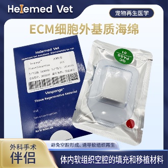
ECM Sponge
ECM filling sponge and ECM cavity-filling biomaterials: for internal soft-tissue cavity filling, providing long-lasting space occupation and inducing regeneration to achieve a soft-tissue graft effect; also an ideal ECM Tumor Filling Material for dead-space repair after tumor resection, and a routine companion to ECM trauma dressings in cavity and fistula wounds.
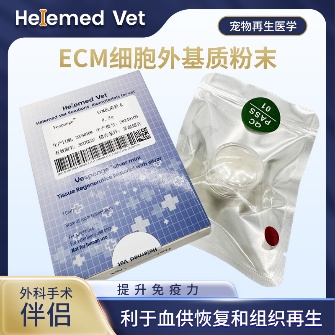
ECM Antibacterial Powder
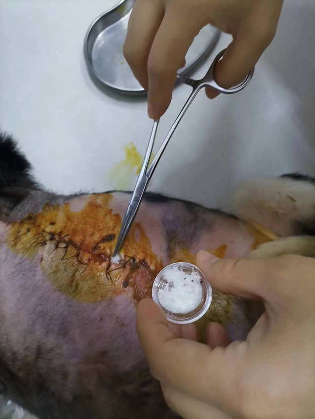
ECM bioscaffold powder — an extracellular matrix (ECM) based antibacterial and inhibitory powder: widely used as ECM trauma dressings on infected internal/external wounds, fresh wounds, burns and crush injuries to markedly improve blood supply, accelerate healing, inhibit infection and reduce complications; it is also an ECM hemostatic material that stops capillary bleeding. How to use: after debridement, take a small amount of powder with forceps and apply to the wound surface, moisten with saline (moist wound healing) and bandage; apply once every 5 days — daily debridement is not required.
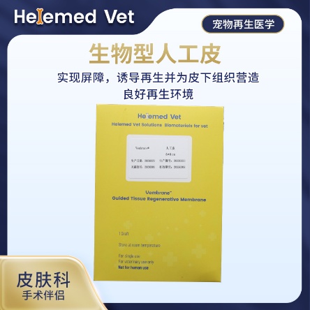
Biosynthetic Artificial Skin (6×8cm)
Artificial skin and ECM trauma dressing for external skin grafts: provides a barrier and induces regeneration, creating a good regenerative environment for subcutaneous tissue in burns and large-area skin loss.
Supplied to veterinary clinics and hospitals. These are surgical materials used by the treating veterinarian as part of a planned procedure — they are not a treatment for a lump, and no material replaces the diagnosis or the removal of the lump itself.
8. Clinical cases
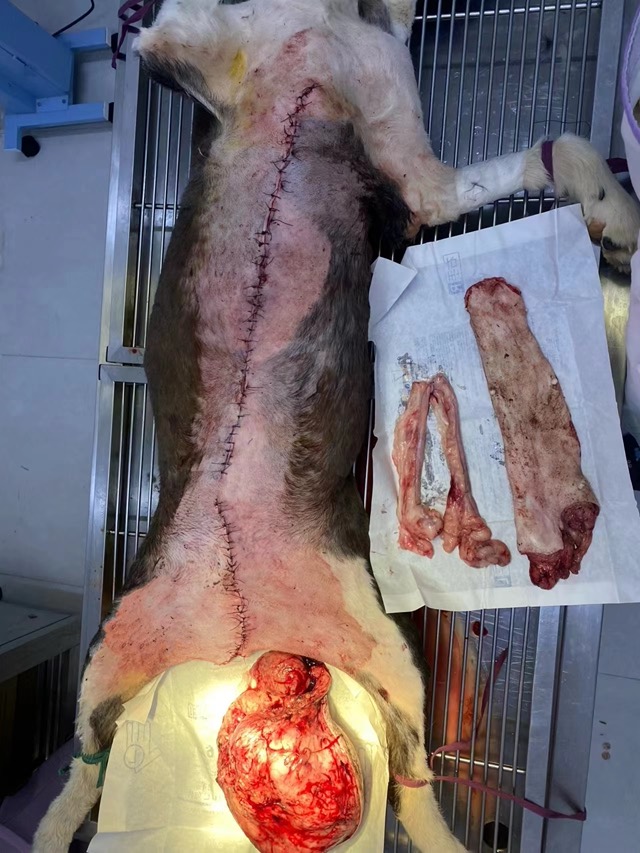
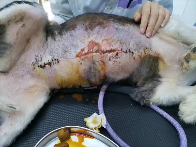
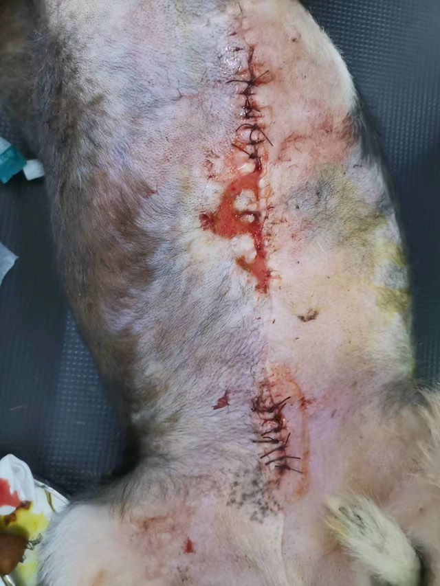
Three cases from our partner hospitals in tumour surgery, where the challenge was not the lump but the space it left: a tumour-resection cavity filled to support healing in an area that heals poorly, a total mastectomy where the powder was laid prophylactically because of the complication rate, and a chest wound that had not healed thirty days after a mastectomy and went on to close.
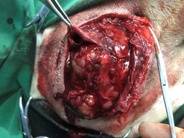
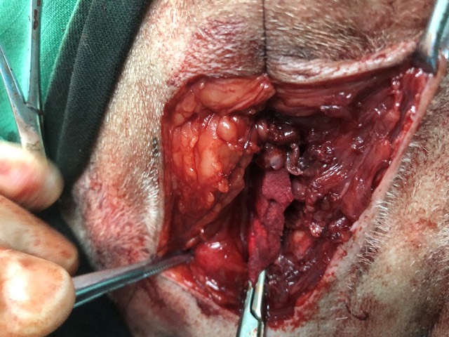
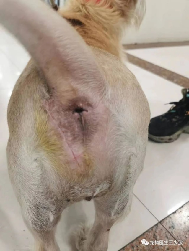
Tumor-Resection Cavity Filling
Tumor-resection surgery often has poor post-op healing; where an obvious cavity exists, implant ECM Sponge for filling (like tissue grafting), lay ECM powder subcutaneously for prophylaxis, then close the wound — effectively accelerating primary healing. Two pieces of ECM Sponge were implanted in this case.
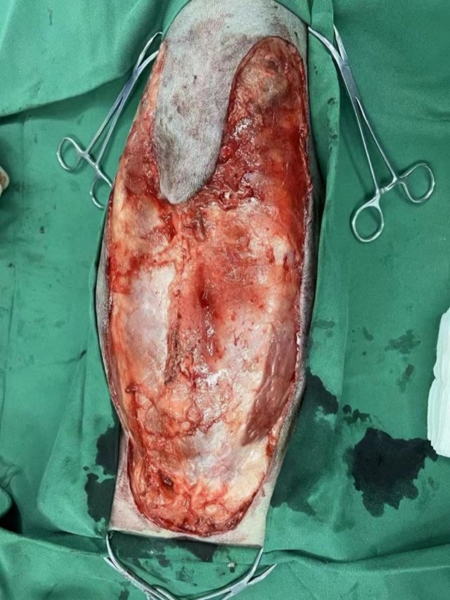
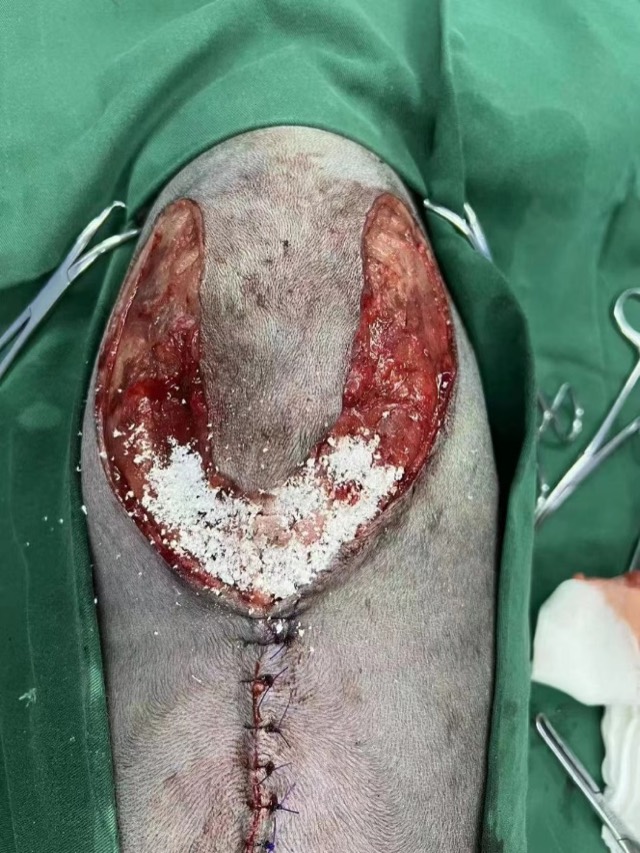
Prophylactic Total Mastectomy Application
The complication rate of total mastectomy is second only to bite cases, so prophylactic application is especially recommended: lay a thin layer of ECM powder subcutaneously in the triangular, high-tension area; use ECM Sponge prophylactically in the inguinal region to avoid cavities and accelerate tissue regeneration.
Non-Healing Wound after Mastectomy
Thirty days after mastectomy the chest wound would not heal; ECM powder was applied subcutaneously and on the surface then bandaged. After 5 days clear signs of regeneration appeared, and it basically recovered after two applications (10 days).
Cases from partner veterinary hospitals. ECM materials are veterinary surgical materials and are applied by the treating veterinarian. See all 39 clinical cases →
Frequently asked questions
How can I tell if my dog’s lump is cancer?
You cannot tell by looking or feeling, and neither can anyone else — the appearance of malignant and benign lumps overlaps too much. What settles the question is a fine needle aspirate, which takes seconds and shows what the lump is made of, or a biopsy if the aspirate is inconclusive. Signs that mean see a vet now rather than later: it is growing week to week, it has broken open or is bleeding, it has become firm or fixed to the tissue underneath, or it is new and growing in an older dog.
Does a fatty lump on a dog need to be removed?
Usually not, once it has been confirmed as a lipoma. Benign fatty lumps in older dogs are common and are generally left alone unless they are large enough to interfere with movement, sit where the dog cannot lie comfortably, or are still growing. The important word is "confirmed" — a soft, movable lump feels reassuring, and reassurance is exactly what makes some serious lumps get missed.
My dog suddenly has a soft lump — is it an abscess?
It may be. An abscess appears over one to three days, is warm and painful to touch, often follows a bite, puncture or foreign body, and may be fluid-filled in the middle. But a cyst, a seroma and some tumours can feel similar. Do not squeeze or lance it at home — pressure pushes infection deeper and risks spreading it, and if the swelling is not an abscess you have made a tumour or cyst worse. Have it examined.
What size of lump should I worry about?
Size on its own is less important than behaviour: how fast it is growing, whether it is fixed to the tissue beneath, whether the surface is breaking down, and how long it has been there. A small lump that can still be removed completely with clean margins is the most curable problem in tumour surgery, so the useful comparison is not "big or small" but "now, when it is simple, or later, when it is not".
Why is my dog’s wound not healing after a lump was removed?
The commonest reason is the cavity left behind rather than the incision. Once a lump is removed, the space between skin and muscle fills with fluid, and that seroma stretches the wound and slows healing. Tension across the closure after a wide removal is the other main cause of a wound opening. Both are managed at the time of surgery, so if a wound is not closing, have it re-examined rather than waiting for it to improve on its own.
Should a mammary lump in my dog be removed straight away?
Mammary lumps should be assessed promptly, because around half are malignant and the prognosis is closely tied to how large the lump is when it is removed. Most are treated by surgical removal, ranging from a single lump to the whole chain on one or both sides depending on how many there are. Before surgery the vet will usually image the chest and sample the local lymph node to check for spread.
Can a dog live normally after a large lump or mastectomy removal?
Yes. The wound is longer and the recovery slower than for a small lump, and there is a period of restricted activity and wound checks, but most dogs return to normal activity once the site has healed. The reason these operations are taken seriously is the weeks immediately after them — the fluid, the tension and the dressings — not the long-term function, which is usually good.