
1. What a hernia actually is
Every hernia has the same three parts, and knowing them makes the whole subject easier to follow:
- The ring or defect — the hole in the wall. It may be present from birth (congenital) or appear later (acquired), and it may be small and firm or wide and floppy.
- The sac — the thin membrane, usually peritoneum, that lines the defect and follows the contents through it.
- The contents — what has slipped through: fat, intestine, bladder, stomach, liver, uterus, or in a spinal hernia, the spinal cord.
A reducible hernia can be gently pushed back into place. An irreducible or incarcerated hernia cannot, usually because the ring is tight or the contents have adhesions. A strangulated hernia is the emergency: the ring has tightened around the contents and cut off their blood supply. Tissue without blood supply dies within hours, and the dog becomes seriously ill.
The single most important thing to understand about a dog hernia: a hernia is defined by the defect, not by the swelling. Pushing the contents back in — if you even can — does not close the hole, and the same contents will push out again. This is why a hernia that keeps reappearing is a surgical problem even when the dog seems perfectly comfortable.
2. The types of hernia in dogs
| Type | Where | Who typically gets it | Urgency |
|---|---|---|---|
| Umbilical | At the navel, through the abdominal wall | Congenital; common in puppies, more in some breeds | Usually elective; emergency if it strangulates |
| Inguinal | In the groin crease | Middle-aged to older females, small breeds; can be congenital | Elective, but the risk of the uterus or bladder becoming trapped is real |
| Perineal | Beside the anus, through the pelvic diaphragm | Older, entire male dogs; the classic "the dog cannot pass stool" case | Urgent but not usually a same-hour emergency; recurrence rate is high |
| Diaphragmatic | Through the diaphragm, between chest and abdomen | Almost always trauma — a road accident or a fall | Emergency. The organs compress the lungs and heart |
| Hiatal | Through the oesophageal opening of the diaphragm | Congenital in some breeds (Shar-Pei, Bulldog); can be acquired | Not usually an emergency; managed medically first in many cases |
| Incisional | Through a previous surgical scar | After abdominal surgery; more likely with poor healing, infection or activity too soon | Elective if small; urgent if the intestine is involved |
In dogs, a perineal hernia and a diaphragmatic hernia are the two that most often end up in surgery, and both are the ones where repair is technically difficult — which is exactly why reinforcement with a mesh is considered in these two.
3. Which hernias are emergencies
Two situations mean "go now, do not wait for Monday":
- Any hernia in a dog that is struggling to breathe. A diaphragmatic hernia puts abdominal organs against the lungs, and the dog cannot breathe deeply. Signs include fast shallow breathing, breathing with the elbows out, blue or pale gums, restlessness, and collapse. This is a genuine emergency.
- Any hernia in a dog that is vomiting, in severe pain, or deteriorating. These are the signs of strangulation — the blood supply to the trapped tissue is cut off. The abdomen may be tense and painful, the dog may vomit repeatedly, and it may go into shock. Strangulated intestine dies, leaks, and causes peritonitis; the difference between surgery on time and surgery late is often whether the dog survives.
Signs of a strangulated hernia in a dog, in the order they usually appear: a soft lump that becomes firm and painful · the dog resents being touched there · vomiting · refusing food · weakness and collapse · pale gums and a racing heart.
What is not an emergency: a small, soft, reducible umbilical hernia in a happy puppy that is eating and playing normally. That one gets an appointment, not a night in intensive care.
4. Signs to look for
- A soft swelling under the skin — at the navel, in the groin, beside the anus, or along an old surgical scar
- A swelling that changes size with position, crying, straining, or after a meal, and that may flatten when the dog lies on its back
- A swelling that feels like it contains something — soft and doughy, or gurgling and intestine-like, or firm like a bladder
- Straining to pass stool, a narrowing of the stool, or a bulge beside the anus that appears when the dog squats — the classic perineal hernia picture
- Difficulty urinating, or blood in the urine, if the bladder has slipped into the defect
- Breathing difficulty after trauma — always ask for a chest X-ray in any dog that has been hit by a car
- Vomiting, pain, loss of appetite or collapse — these mean the hernia may be strangulated
Two practical notes. First, an umbilical hernia is often found in a puppy at the first vaccination visit and is not the same thing as an umbilical infection, which is a moist red stump in a newborn and is treated very differently. Second, not every lump is a hernia — lipomas, abscesses, tumours and enlarged lymph nodes all present as lumps, and a vet distinguishes them by feel and, where needed, by ultrasound or a needle sample.
5. How a hernia is diagnosed
- Palpation — the vet feels the defect and its edges, checks whether the contents can be reduced, and judges how big the ring is. This is done gently: squeezing a hernia can damage the contents.
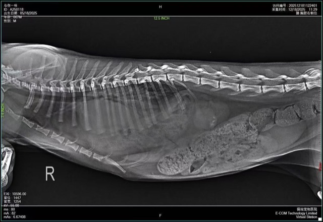
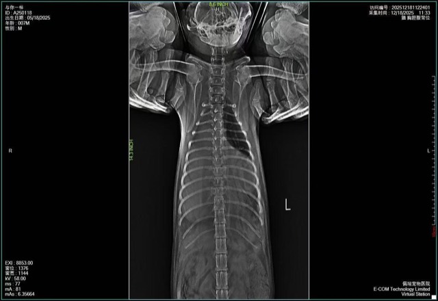
- Radiography — the key test for a diaphragmatic hernia, where the X-ray shows abdominal organs in the chest and a loss of the normal diaphragm line.
- Ultrasound — identifies what is inside the sac (fat, intestine, bladder, uterus) and whether the tissue still has a blood supply. This changes the urgency and the surgical plan.
- Contrast studies — a barium swallow for a suspected hiatal hernia, or contrast into the bladder when a urinary tract defect is suspected.
- Blood work and electrolytes — a strangulated hernia causes metabolic changes that must be corrected before anaesthesia.
In an unstable trauma patient, the order is reversed: the dog is stabilised, the chest is assessed for bleeding and lung contusion, and surgery for the hernia may be delayed until the dog can survive the anaesthetic — unless the hernia itself is what is preventing the dog from breathing.
6. How hernias are repaired
Almost all hernias that need treating are repaired surgically, and all repairs follow the same logic: return the contents, remove the sac, and close or reinforce the defect.
- Reduce the contents. The organs are returned to where they belong. If the tissue is dead — dark, cold, without bleeding at the cut edge — it is resected.
- Reconstruct the wall. A small defect with healthy tissue is closed by suturing the edges together. But the wall must close without tension: sutures under tension cut through tissue, which is how a repair fails.
- Reinforce when the tissue cannot hold. Where the defect is large, where the surrounding tissue is thin or degenerated, or where the anatomy means the repair will always be under load, a reinforcing mesh is implanted and sutured to the surrounding tissue.
- Drain and close. Depending on the site and the amount of dead space, the surgeon may place a drain. The skin is closed over the repair.
Age and general health change the plan more than the hernia does. An older male dog with a perineal hernia may have a heart murmur, kidney values that are not ideal, and a prostate problem that is contributing — all of which are addressed in the same admission.
And a note on the thing owners ask most often: no, a hernia does not heal by itself. The defect is a structural hole in a wall. It may close in a very young puppy with a small umbilical defect, and that is the one exception worth a wait-and-see period, but the defect does not disappear in an adult dog.
7. When a mesh is needed
The word "mesh" worries owners, who picture a permanent piece of plastic left in the dog. In dogs, the mesh used for hernia repair is typically a biosynthetic, absorbable scaffold rather than a permanent synthetic screen — a decellularised biological matrix that the dog’s own cells and blood vessels grow into, rebuilding the region and then being replaced by the animal’s own tissue.
Mesh is considered when suturing the tissue alone is likely to fail:
- Large defects where the edges cannot be brought together without tension
- Thin or degenerated tissue, which is the norm in older dogs with perineal hernias
- Diaphragmatic and thoracic defects, where the repair is under constant movement from breathing
- Revision of a failed repair — the first attempt already showed the tissue will not hold
- Sites where recurrence is common, which is why a perineal hernia repair is often reinforced from the start
An absorbable mesh has two practical consequences worth understanding. It has to be sutured under appropriate tension, without being stretched tight, because it must stay in position long enough for tissue to grow in; and because it degrades, drainage is recommended — the fluid that collects around a degrading implant has to go somewhere, and a seroma left to build up can become an infection.
A permanent foreign material left in a body under load, in an area that is never completely sterile, eventually becomes a chronic problem. That is the argument for a scaffold that is replaced by the dog’s own tissue rather than a screen that stays forever.
Materials used in hernia repair
Our ECM regenerative materials are veterinary surgical materials used by the treating veterinarian. The biosynthetic absorbable hernia mesh is a biological hernia patch and an absorbable hernia patch in one implant, used for internal hernia surgery and thoracic repair; it is sutured to the surrounding tissue after the hernia ring is closed or reduced, and drainage is recommended.
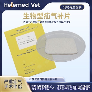
Biosynthetic Hernia Mesh (5×6cm)
Biosynthetic Absorbable Hernia Mesh — a biological hernia patch and absorbable hernia patch in one implant: for internal hernia surgery and thoracic repair; biodegradable, fully rebuilding the hernia region without long-term foreign-body sensation; drainage is recommended.
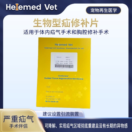
Biosynthetic Hernia Mesh (7×9cm)
Large-size biological hernia patch / absorbable hernia patch: for large or recurrent hernias, internal hernia surgery and thoracic repair, full reconstruction with no long-term foreign-body sensation.
Available in two sizes — 50×60mm for smaller rings and 70×90mm where the defect is wide or needs to be bridged. Supplied to veterinary clinics and hospitals.
8. Recovery after hernia surgery
The repair is only half of it. What happens in the first four weeks decides whether the hernia comes back:
- Activity restriction, strictly. No jumping, no stairs, no running, no playing with other dogs. Often four to six weeks. Increased pressure inside the abdomen — from jumping, straining or coughing — is what pushes a repair apart.
- Drain and wound care. Keep the site clean and dry, check the drain output as instructed, and report any swelling, discharge or smell.
- Stool softeners and diet. After a perineal hernia repair, straining to pass stool is the main threat to the repair. Soft stool, a high-fibre or easily digested diet, and sometimes a stool softener are part of the treatment.
- A collar. Licking introduces infection into a fresh repair.
- Recheck appointments. Recurrence is detected by feel, often before the owner notices anything.
Recurrence is a real possibility, and it is highest for perineal hernias — the pelvic diaphragm in these dogs is generally degenerated, not just torn in one spot. Hormonal status matters too, which is why neutering is usually discussed at the same time. A second repair is often reinforced with a mesh even if the first was not.
9. Clinical cases
Three cases from our partner hospitals: a perineal hernia repaired with the biosynthetic mesh, a diaphragmatic hernia where abdominal organs were filling the chest, and the animal experiment and pathological validation that show what happens inside the mesh after implantation.
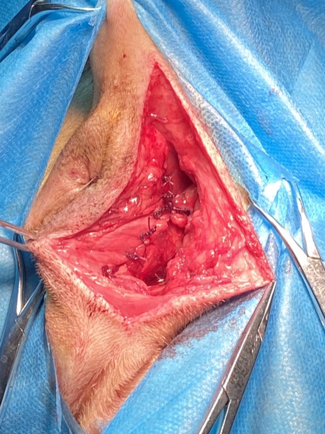
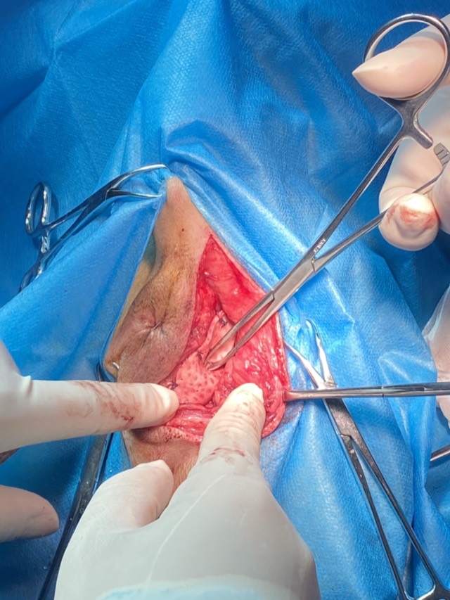
Perineal Hernia Repair
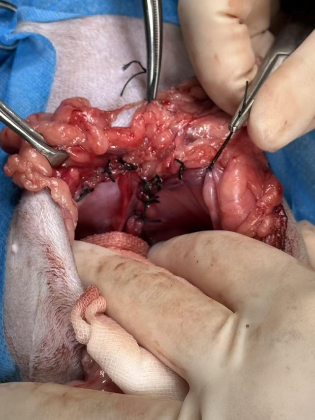
The only domestic biosynthetic hernia mesh (porcine decellularized process): first close or minimize the hernia ring, then suture the mesh to the surrounding healthy tissue; its natural pores make needle entry easy without destroying physiological structure. Early support shares tension; later cells and vessels grow in for full reconstruction — biodegradable with no long-term foreign-body sensation.
Diaphragmatic Hernia Repair
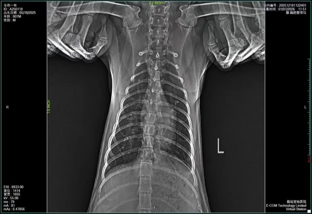
X-ray showed abundant abdominal-organ shadows in the thoracic cavity with compressed heart and lungs, confirming diaphragmatic hernia: large-area defect and rupture of the diaphragm. The defect was precisely measured and a custom-cut, slightly larger biosynthetic mesh fully covered and repaired it; X-ray at 12 days post-op showed good recovery and the patient was discharged.
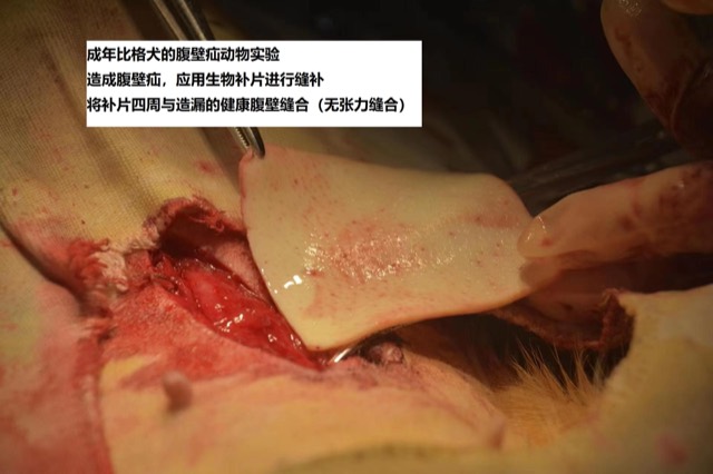
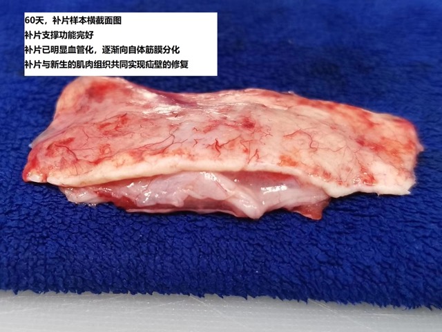
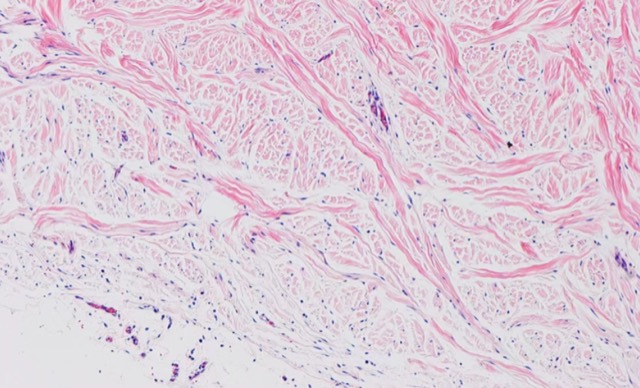
Hernia Mesh Animal Experiment & Pathological Validation
Biosynthetic Absorbable Hernia Mesh animal experiment: after implantation, cells and vessels grew in, fully rebuilding the hernia region; the 60-day pathological section showed fibroblasts infiltrating the mesh with abundant differentiated mature fibrocytes and vascular distribution, and no inflammatory reaction.
Cases from partner veterinary hospitals and approved animal research. ECM materials are veterinary surgical materials and are applied by the treating veterinarian. See all 39 clinical cases →
Frequently asked questions
Is a dog hernia painful?
A simple, reducible hernia is usually not painful — owners often find a soft lump that the dog does not mind being touched. A hernia becomes painful when the contents are trapped: the swelling turns firm, the dog resents handling, and it may vomit or go off its food. That change is the signal to go to a vet immediately.
Can a dog hernia heal on its own?
A small umbilical hernia in a very young puppy sometimes closes as the puppy grows, and that is worth watching for a few months. Apart from that exception, a hernia is a structural hole in a wall and does not close by itself. A soft lump that can be pushed back in still has a hole behind it, and the contents will push out again.
How do I know if my dog’s hernia is strangulated?
The lump becomes firm, tense and painful; the dog may vomit repeatedly, refuse food, become weak or collapse, and the gums may be pale. Strangulation cuts off the blood supply to trapped tissue, so this is a same-hour emergency — not something to watch overnight.
What are the signs of a hernia in a dog after being hit by a car?
Fast, shallow or laboured breathing, breathing with the elbows pushed out, restlessness, pale gums, and a belly that looks different from normal. Any dog that has been hit by a car should have its chest imaged, because a diaphragmatic hernia and lung bruising are common and can be present even when the dog walks away from the accident.
Why does my older male dog strain to pass stool and have a swelling beside the anus?
That is the classic presentation of a perineal hernia — the pelvic diaphragm has given way and the rectum is bulging into the space beside it. It needs a veterinary examination; the longer it is left, the higher the chance the bladder or intestine becomes trapped, and the harder the repair becomes.
Why is a mesh used instead of just stitching the hernia closed?
Because sutures under tension cut through tissue over time, which is the most common way a repair fails. A mesh spreads the load across the whole defect, which matters most in large defects, in older dogs whose tissue is thin, and in repairs under constant movement such as the diaphragm.
Does the hernia mesh stay in the dog forever?
The biosynthetic mesh we supply is absorbable. It is a scaffold: the dog’s own cells and blood vessels grow into it and rebuild the area, and the implant degrades and is replaced by the dog’s own tissue. Because it degrades, drainage is recommended after implantation to remove the fluid that collects around it.