
- 1. What counts as a fracture
- 2. Signs your dog has a fracture
- 3. First aid in the first 30 minutes
- 4. How a fracture is diagnosed
- 5. How each type is treated
- 6. The bone healing timeline
- 7. Why some fractures never unite
- 8. Bone defects and revision surgery
- 9. Clinical cases
- Frequently asked questions
- Related guides
1. What counts as a fracture
"Broken bone" and "fracture" mean the same thing — a fracture is simply the medical word for a break. What matters clinically is the type, because the type decides the treatment:
| Type | What it means | Typical treatment |
|---|---|---|
| Closed (simple) | The bone is broken but the skin is intact. | Depends on the bone; often surgery, but some can be splinted |
| Open (compound) | The bone has broken through the skin, or there is a wound reaching the fracture. | Emergency surgery. Infection risk is high — this is the single most important thing to distinguish |
| Incomplete / greenstick | The bone is cracked or bent, not fully separated. Common in puppies because their bones are softer. | Usually a splint or strict rest, but it must be re-checked — a greenstick fracture can complete |
| Comminuted | The bone is shattered into multiple fragments. | Surgery. Needs a plate, pins or an external fixator, and often a bone graft to fill the gaps |
| Articular | The break enters a joint surface. | Surgery. Joint surfaces must be reconstructed accurately or arthritis follows |
| Pathological | The bone broke through a weak point — a bone tumour, a cyst, or severe bone loss from infection. | Treat the cause, not just the break |
Two distinctions change everything about the next hour. Open versus closed decides whether the dog needs antibiotics and surgery tonight. And which bone decides how serious it is: a fractured toe is uncomfortable, while a fractured femur, pelvis or spine can be life-threatening because of the blood loss, internal injury and nerve damage that come with it.
Suspected spinal or pelvic fracture: do not lift the dog normally. Keep the body straight, slide a rigid board or firm mattress under it, and carry it flat. Twisting the spine can turn a recoverable injury into permanent paralysis.
2. Signs your dog has a fracture
The obvious ones are easy. The subtle ones are where owners get caught out, especially with incomplete fractures in puppies:
- Not putting weight on a leg — or touching the toe to the ground and immediately lifting it
- Swelling at the site, which appears within minutes to hours and keeps growing
- Obvious deformity or an unnatural angle — a limb that visibly does not match the other side
- Bone grating (crepitus) if the limb is moved — never test this deliberately
- Crying, panting, trembling, or a change in behaviour that suggests severe pain
- An open wound over a bone, sometimes with a piece of bone visible — this is an open fracture until proven otherwise
- Sudden reluctance to jump, climb stairs or be picked up — with spinal or pelvic fractures this can be the only sign
- Dragging a limb, or a limb that feels floppy or has no pain response — a possible nerve injury, which must be assessed urgently
What it is often confused with: a torn cruciate ligament (sudden hind-limb lameness with no deformity, very common in larger dogs), a luxated knee (patella), a muscle strain, or a penetrating foreign body in the pad. All of these need the same first step — a veterinary examination — because you cannot tell them apart from the outside.
3. First aid in the first 30 minutes
- Stop the dog moving. Confine it in a crate, a box or a carry bag. Movement is what turns a closed fracture into an open one and what makes internal bleeding worse. If the dog is struggling, cover the head with a light towel to reduce visual stimulation — do not sedate it yourself.
- Control bleeding first. For an open fracture or a bleeding wound, apply firm direct pressure with a clean pad. If blood soaks through, add another pad on top rather than removing the first one.
- Do not try to set or straighten the bone. Pulling on a limb to "line it up" can sever a blood vessel or nerve and can push a closed fracture through the skin. Leave the limb in the position you found it.
- Support, do not splint. You can support the limb against the body with a folded towel or a soft bandage for transport, but do not apply a rigid home-made splint. A splint that is too tight causes swelling and tissue death, and a splint that is too loose does nothing. Immobilise the whole dog, not the bone.
- Muzzle if needed, even a friendly dog. A dog in severe pain can bite the person it loves. Use a soft muzzle or a strip of bandage over the nose — but never muzzle a dog that is vomiting or having trouble breathing.
- Cover an open fracture with a clean, damp dressing — sterile saline-soaked gauze if you have it, a clean cloth if you do not. Do not flush a deep wound with disinfectant, alcohol or hydrogen peroxide, and do not push bone fragments back in.
- Call the clinic before you set off, so surgery and radiography are ready when you arrive, and describe: which limb, open or closed, bleeding yes or no, can the dog feel its toes, is it breathing normally.
Do not give human painkillers. Paracetamol (acetaminophen) and ibuprofen are toxic to dogs and cause liver failure, kidney failure and stomach ulceration. In a dog that may need surgery, giving medication by mouth can also delay anaesthesia. Keep the dog warm, keep it still, and let the vet manage the pain.
4. How a fracture is diagnosed
Radiography is the answer, and it is not always as simple as one picture:
- Two views minimum, at right angles, of the whole bone including the joint above and below. A fracture near a joint is easily missed on a single view.
- Sedation or general anaesthesia for the X-ray. A dog in pain will not hold a limb still, and a blurred film hides the fracture and the number of fragments.
- Radiographs of the opposite limb in puppies, so the vet can compare the growth plates and not mistake a normal growth plate for a fracture line.
- Thoracic and abdominal imaging in road-traffic cases, because the chest and abdomen are injured more often than owners expect — pulmonary contusions and internal bleeding kill more dogs than the fracture itself.
- CT for complex, comminuted or spinal fractures, where 3D reconstruction changes the surgical plan.
If the dog is stable, the fracture is closed and the limb is not at risk, the vet may need to stabilise first and image later — shock, breathing difficulty and internal bleeding take priority over the bone.
5. How each type is treated
There is no single answer, because a fracture in a Chihuahua's toe and a fracture in a Great Dane's femur are different problems. The decision comes down to three questions: is the fracture stable or unstable, does it involve a joint, and how much load has the bone to carry.
| Approach | Used for | Notes |
|---|---|---|
| Strict rest and external coaptation (splint, cast, sling) | Stable, incomplete fractures; some pelvis fractures; toe and metacarpal fractures | Cheap and non-invasive, but needs 4–8 weeks of genuine restriction, and the cast must be checked — rubbing casts cause serious pressure sores |
| External fixator (pins through the skin into the bone, joined by a bar outside) | Highly comminuted fractures, open fractures with soft-tissue damage, fractures that are already infected | Does not disturb the fracture site, allowing the wound to be managed; the pin tracts need daily cleaning |
| Internal fixation (bone plate and screws, or pins and wires) | Most femur, tibia, humerus, radius and ulna fractures | The most common approach. Restores the bone to normal anatomy and allows earlier use of the limb |
| Plate or pin plus bone graft | Comminuted fractures with missing fragments, and fractures with a gap | Filling the gap matters: bone does not bridge thin air, and a persistent gap is a common reason a fracture fails to unite |
| Arthrodesis (surgical fusion of a joint) | Fractures that destroy a joint surface beyond reconstruction, usually the carpus or hock | A salvage procedure — it removes the joint, and with it the pain |
| Amputation | Limb damage that cannot be reconstructed — crushed limbs, severe open fractures with dead tissue, or where reconstruction is not affordable | Dogs do remarkably well on three legs; it is a legitimate option, not a failure |
Whichever route is chosen, the wound and the soft tissue around it matter as much as the bone. An open fracture with dead muscle, contamination and no blood supply will not heal whatever plate is put on it, which is why the first surgery is spent on irrigation and debridement.
6. The bone healing timeline
Bone heals in stages, and the stages are predictable even though the calendar is not. In a young dog with a simple fracture of a well-immobilised bone, it can be fast; in an older, large-breed dog with a comminuted or open fracture, expect the upper end of every range.
| Time after surgery | What is happening in the bone | What you will see |
|---|---|---|
| Days 1–7 | Inflammation, a blood clot forms at the fracture line, cells migrate in | Swelling and bruising peak then start to settle; the limb is still very painful; strict confinement |
| Weeks 2–3 | Soft callus forms — a bridge of cartilage and fibrous tissue holds the fragments | Toe-touching may start; stitches come out; the X-ray shows a fuzzy bridge but no solid bone yet |
| Weeks 4–8 | The soft callus mineralises into hard callus — this is where the bone becomes genuinely stable | Increasing weight-bearing; the X-ray shows cloudy new bone across the fracture |
| Months 3–6 | Remodelling: the callus is reshaped into normal structural bone | Near-normal walking; a plate may stay in permanently or be removed once the bone is consolidated |
Two rules keep this on track. Restriction is a treatment, not a suggestion — a dog that is allowed to jump on the sofa at week three can break the fixation, and a second surgery costs more than the first. And the X-ray decides, not the dog: dogs will use a limb long before the bone is strong, which is why the vet re-radiographs at set intervals rather than trusting how the dog looks.
What helps healing: strict restriction, good pain control (pain slows healing), adequate protein and calories, and keeping the dog at a healthy weight. What hinders it: early free exercise, obesity, smoking in the home, malnutrition, infection, and the use of steroids and some anti-inflammatories in the early phase.
7. Why some fractures never unite
When a fracture is still not healed months later, vets call it a delayed union or, if the bone has stopped trying, a non-union. It is not bad luck. There are a small number of recognisable causes, and each has a fix:
- Instability. The most common cause by far. If the fragments still move, the body forms fibrous tissue instead of bone. Fixed by better fixation — a stronger plate, more screws, or a change of technique.
- Infection. Bacteria on the plate keep the fracture inflamed and resorb bone around the implants. This is the "bone dissolution" seen on X-rays after orthopedic surgery, and it is why an infected fracture needs the implants cleaned, replaced or removed and the site debrided.
- Loss of blood supply. A fragment stripped of its periosteum by the injury or by a rough surgery becomes dead bone. Dead bone cannot unite — it has to be removed and the gap filled.
- A gap that was never filled. If a fragment was lost, or a piece of dead bone was removed, the gap stays. Bone will not bridge a gap it cannot reach across.
- Soft tissue interposed. Muscle caught between the fragments stops the bone from touching.
- Systemic factors. Steroid treatment, hypothyroidism, Cushing’s disease, malnutrition, and some painkillers given in the early phase.
The practical consequence: a fracture that has not united needs imaging and usually surgery — not more time. And when the revision is done, the surgeon will often add a graft, because the reason the first attempt failed is usually that the site lacked stability, blood supply, or material to bridge the defect.
8. Bone defects and revision surgery
A bone defect is a gap or a void in the bone. It happens after a comminuted fracture with missing fragments, after dead or infected bone is removed, and after tumour resection. Defects are the hardest part of fracture surgery, because the implant holds the bone in place but cannot by itself make new bone grow across a gap.
Options for filling a defect vary with size and with whether the site is infected:
- Autograft — bone taken from the dog itself, typically the iliac crest. The gold standard for cell viability, but it means a second surgical site, limited quantity, and more pain.
- Allograft — bone from a donor dog. Available in bigger volumes, but no living cells and a small infection risk.
- Bone substitutes and scaffolds — synthetic ceramics, and extracellular matrix (ECM) based implantable materials, which act as a scaffold for the dog’s own cells to migrate into and lay down new bone.
In a site that is or has been infected, the choice narrows, because a plain graft in a contaminated bed tends to become a dead, infected piece of material. This is why materials that combine a scaffold with an antibacterial effect are used in revision surgery — and why the surgeon will usually wait until the infection is under control before placing anything.
The sequence of a typical revision is: remove the failed implants, debride dead and infected bone back to bleeding bone, stabilise the fracture properly, fill the defect, ensure soft tissue covers the site, and culture the tissue so the right antibiotic is used. Take away any one of those steps and the revision fails the same way the first surgery did.
Materials used when a fracture leaves a bone defect
Our ECM regenerative materials are veterinary surgical materials used by the treating veterinarian, as part of a fracture repair protocol and always after debridement. ECM artificial bone fills the defect and acts as a scaffold for new bone, with a sustained antibacterial effect; the larger blocks are used where a limb-bone defect has to be filled.
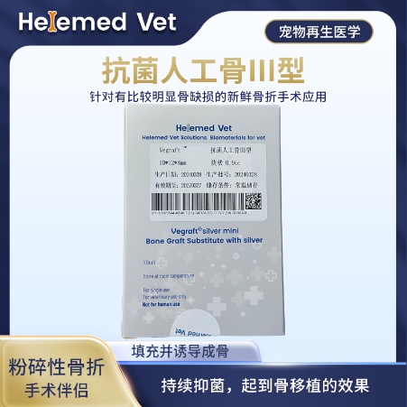
Antibacterial Artificial Bone Type III
Limb-bone ECM regenerative bone material for fresh fracture surgery with obvious bone defects; an ECM implanted bone material that fills the defect and induces osteogenesis with a sustained antibacterial effect, achieving a bone-graft effect.
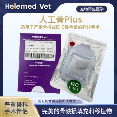
Artificial Bone Plus Type I
ECM bioscaffold and extracellular matrix bone repair material: fills bone defects in old fracture revision surgery, inducing osteogenesis with better activity, for ECM regenerative bone material and bone-graft procedures.
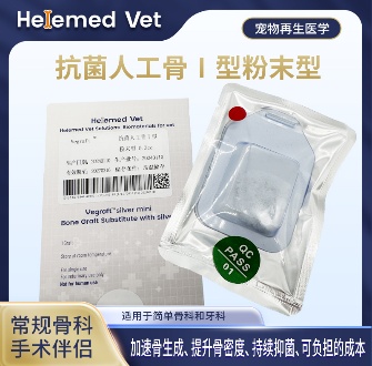
Antibacterial Artificial Bone Type I (Powder)
ECM artificial bone material in powder form — an ECM regenerative bone material and ECM bone repair material: for fresh fracture surgery and periodontal GTR / GBR guided regeneration, inducing osteogenesis and providing a sustained antibacterial effect.
These materials are supplied to veterinary clinics and hospitals. Ask your veterinarian whether they fit your dog’s fracture and whether the site is suitable for grafting.
9. Clinical cases
Three cases from our partner hospitals, all of them fractures that had already failed once: a tibial fracture that re-fractured after the plate was removed, an old comminuted fracture with a very large bone defect on its third revision, and a fracture fixed with a graft, followed on X-ray.
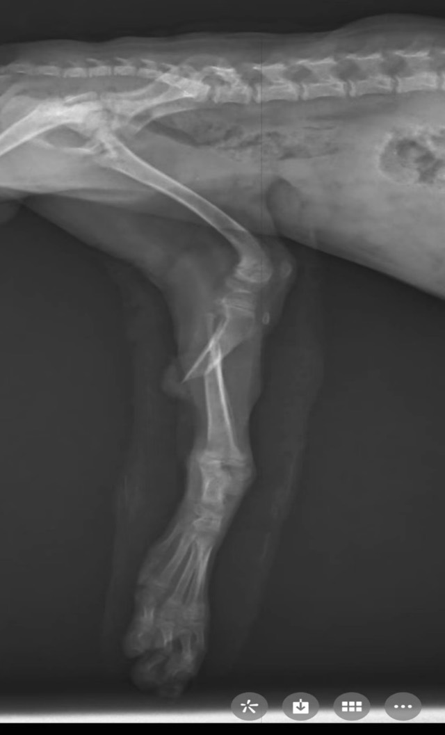
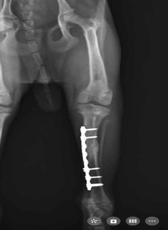
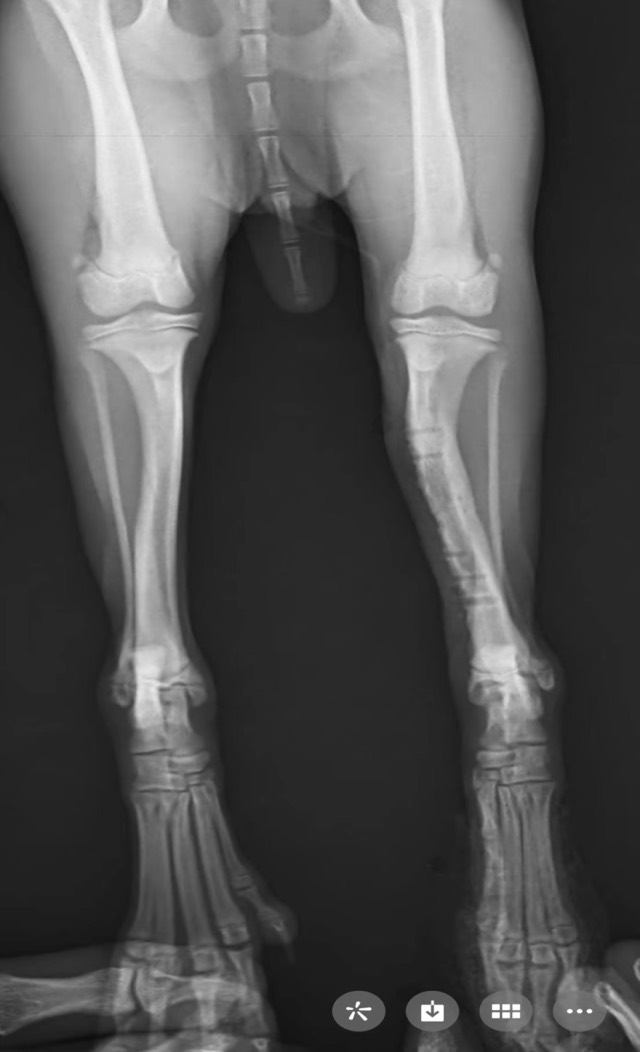
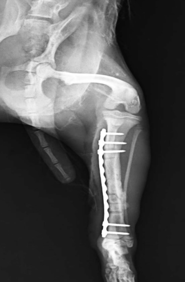
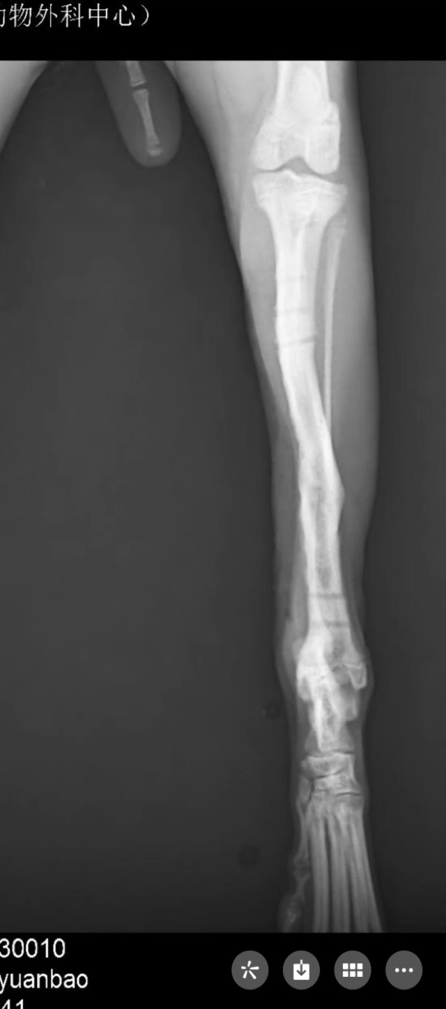
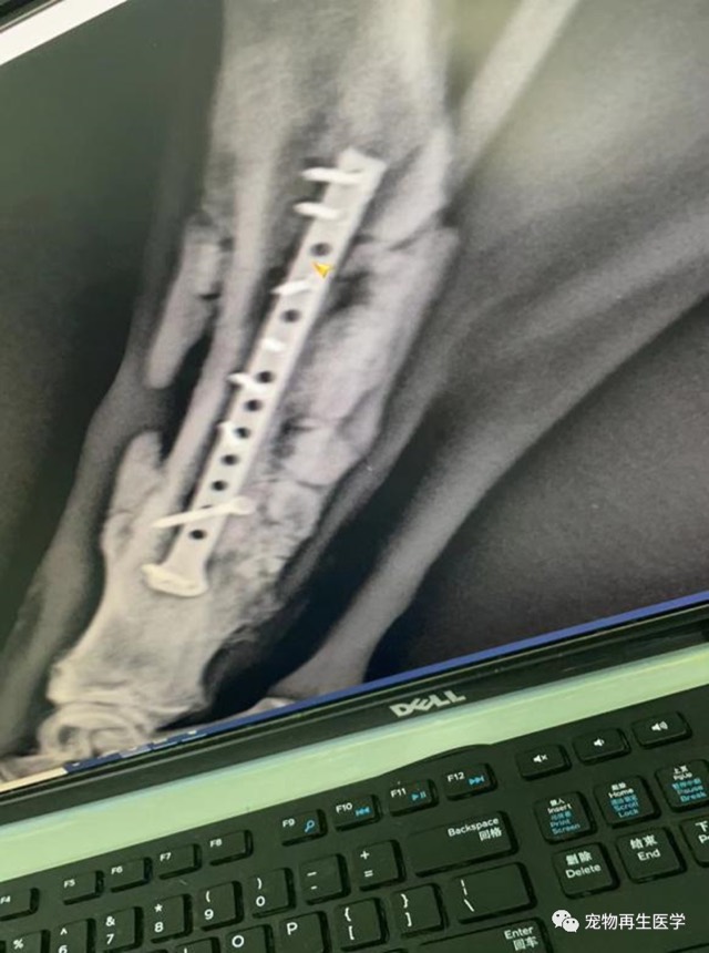
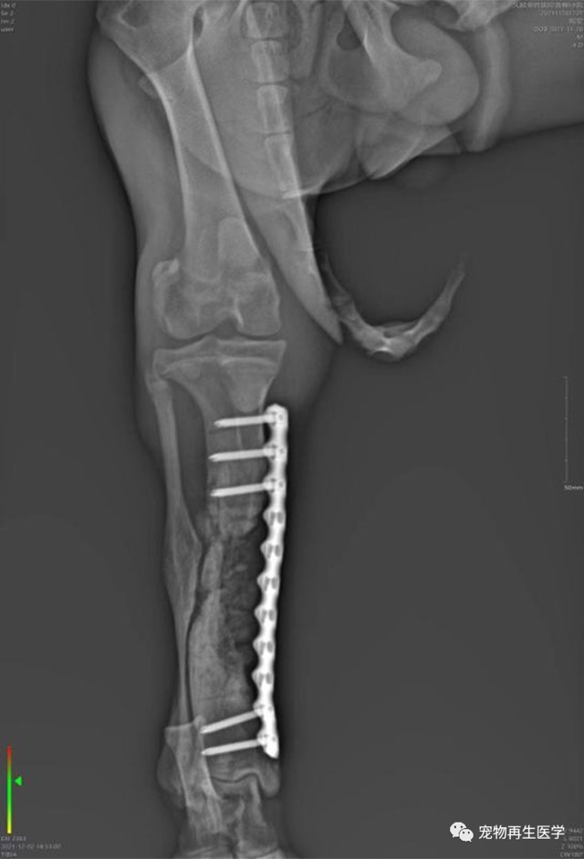
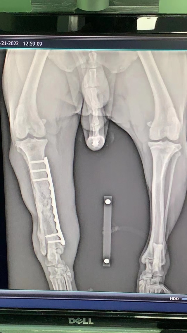
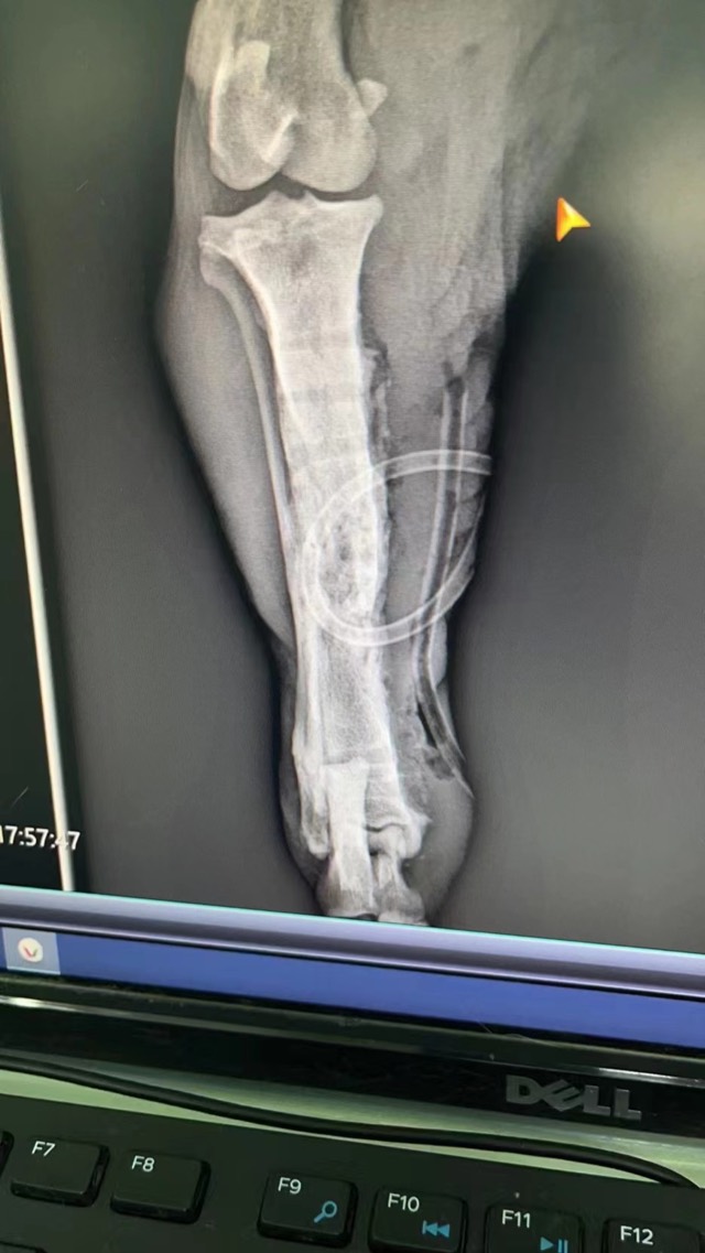
Tibial Oblique Fracture Re-Fracture Revision
After the first internal fixation of a tibial oblique fracture and plate removal following normal recovery, a secondary fracture appeared at the third screw hole one week later (common in small animals). The second fixation used Antibacterial Artificial Bone granules — filling the defect where present, or around the fracture line where not; the new bone density was markedly higher. At plate removal after healing, Antibacterial Artificial Bone was packed into the screw holes to accelerate osteogenesis, and no further secondary fracture occurred.
Old Comminuted Fracture Revision
A third revision surgery for severe comminuted, infected fracture with very large bone defect; Artificial Bone Plus (ECM bioscaffold with added BMP) was used together with a periosteal patch — isolating soft tissue to prevent premature invasion and forming early periosteum through vascularization to nourish osteogenesis. Eventually the plate was removed and movement recovered as before.
Fracture Fixation and Bone Grafting (X-ray Follow-up)
Bio-artificial bone (ECM + bioactive ceramics) was used for fracture fixation and bone grafting; X-ray follow-up showed new bone formation and increased bone density.
Cases from partner veterinary hospitals. ECM materials are veterinary surgical materials and are applied by the treating veterinarian. See all 39 clinical cases →
Frequently asked questions
How long does a dog fracture take to heal?
A simple fracture in a young dog can be stable in four to six weeks; a comminuted or open fracture in an older large-breed dog commonly takes three to four months, sometimes longer. The timeline is driven by the bone involved, the dog’s age and size, the stability of the fixation and whether infection is present. Your vet will confirm healing on X-ray, not by how the dog looks.
Can a dog fracture heal without surgery?
Sometimes. Stable, incomplete fractures and some pelvis and toe fractures are treated with strict rest and external support. Unstable fractures, fractures that enter a joint, and most femur and tibia fractures will not heal reliably without surgery — and a limb that heals in the wrong position causes lifelong arthritis.
What should I do right now if I think my dog has a broken leg?
Keep the dog still, support the limb without trying to straighten or splint it, muzzle if there is any chance of being bitten, cover an open wound with a clean damp dressing, and go to a veterinary hospital. Do not give human painkillers. Call ahead so the clinic is ready.
My dog had fracture surgery and the bone is not healing. Why?
The usual causes are instability of the fixation, infection around the implant, dead bone with no blood supply, an unfilled gap between fragments, or soft tissue trapped in the fracture. All of them need investigation with new X-rays and usually further surgery — time alone does not fix them.
What is bone dissolution after orthopedic surgery?
It is the loss of bone around the implants, visible on X-ray as a dark zone where mineral has disappeared. It is typically driven by infection or by micromovement of a loose implant, and it weakens the fixation. It is treated by identifying the organism, controlling the infection and revising the fixation.
Is a bone graft always needed for a dog fracture?
No. A simple fracture that lines up and is stably fixed will fill itself in. A graft or a bone substitute is needed when there is a gap to bridge — missing fragments, dead bone that had to be removed, a non-union being revised, or a defect after tumour surgery.
Will the plate have to be removed later?
Often it can stay for life, particularly in older dogs. Removal is considered when the implant causes discomfort, becomes infected, loosens, interferes with a growing dog, or once the bone is fully consolidated and the owner prefers it out. Removal is a second surgery with its own recovery, so it is a decision, not a routine.