
- 1. What a skin infection actually is
- 2. The four types, and how they differ
- 3. Signs, and where they appear
- 4. Why some dogs keep getting them
- 5. How a vet diagnoses it
- 6. How it is treated
- 7. Why antibiotics alone often fail
- 8. When the skin breaks down
- 9. Clinical cases
- Frequently asked questions
- Related guides
1. What a skin infection actually is
Healthy dog skin carries a permanent population of bacteria and yeast — mostly Staphylococcus pseudintermedius and Malassezia pachydermatis. They cause no trouble while the skin barrier is intact and the immune system is normal. A skin infection happens when one of those two things changes:
- The barrier breaks. Scratching, chewing, a hot spot, a wound, a foreign body, excessive washing, or a coat that stays damp.
- The skin's defences drop. Allergy, hormonal disease, poor nutrition, or immunosuppressive medication.
This is why so few skin infections are "just an infection". In the great majority of recurrent cases, the infection is the visible symptom of an underlying skin disease, and the underlying disease is what has to be managed.
2. The four types of dog skin infection
| Type | What it looks like | Itch level | Confirmed by |
|---|---|---|---|
| Bacterial — superficial pyoderma | Papules, pustules, circular scaling "collarettes", crusts, patches of hair loss. Short-coated dogs show moth-eaten bald patches. | Moderate to severe | Cytology (stained smear) and tape preparation |
| Bacterial — deep pyoderma | Nodules, draining tracts, ulcers, swelling, sometimes fever. Often on the chin, between the toes, or pressure points. | Severe, painful | Cytology plus bacterial culture |
| Yeast — Malassezia | Greasy, thickened, musty-smelling skin, often dark and leathery. Chronic ear infection is usually part of it. Classic sites: armpits, groin, neck, feet. | Severe | Cytology (tape impression) |
| Fungal — ringworm (dermatophytosis) | Circular bald patches with a scaly, sometimes reddened rim; broken hairs. Slow-moving. | Mild | Wood's lamp, fungal culture, PCR |
| Parasitic — demodectic or sarcoptic mange | Patchy hair loss and scaling, often around the eyes and muzzle (Demodex) or generalised intense itch with crusting (Sarcoptes). | Mild to severe | Deep skin scrape |
Two practical points. First, lesions frequently overlap: a dog with flea allergy often has both a bacterial and a yeast infection at the same time, and treating only one of them is why the dog never quite gets better. Second, ringworm and sarcoptic mange are contagious to people and other pets, so they change the whole household plan — they are worth ruling out early rather than after two months of shampoo.
3. Signs, and where they appear
- Redness, papules, pustules, crusts, scales, or greasy odour from the coat
- Hair loss in patches, especially the classic "moth-eaten" look on the trunk of short-coated dogs
- Thickened, dark, elephant-like skin in chronic cases
- Constant licking, chewing, rubbing, scooting, or head shaking
- Bad smell — a musty, yeasty odour strongly suggests Malassezia
- Pain when touched, or the dog not wanting to be handled
- Draining tracts or nodules → deep infection, which needs more than shampoo
| Where it is | Think first of |
|---|---|
| Lower back, tail base, hindquarters | Flea allergy dermatitis with secondary pyoderma |
| Face, muzzle, chin ("stud tail" / chin acne) | Contact or plastic-bowl dermatitis, Demodex, food allergy |
| Ears, especially if recurrent | Malassezia otitis, often with underlying allergy |
| Armpits, groin, neck folds, feet | Atopy with secondary bacterial and yeast infection |
| Between the toes, nail folds | Foreign body, Demodex, occasionally deep pyoderma or fungal infection |
| Anywhere with a circular scaly rim | Ringworm — treat as contagious until proven otherwise |
4. Why some dogs keep getting skin infections
A dog that has had three skin infections in a year does not have "bad luck". Something is making the skin vulnerable, and it is usually one of these:
- Environmental allergy (atopy) — the single most common underlying cause of recurrent pyoderma
- Flea allergy dermatitis — one flea bite drives an intense, self-traumatising itch
- Food allergy — less common, but a real cause of non-seasonal itch and recurrent ear disease
- Hypothyroidism — thickened skin, symmetrical hair loss, recurrent infection, weight gain, low energy
- Cushing's disease — thin skin, pot belly, recurrent infection, increased drinking
- Poor nutrition or low protein, which slows skin repair
- Immunosuppressive medication, including long steroid courses
- Anatomy — skin folds in Bulldogs, Pugs and Shar-Peis trap moisture and heat
If your vet has diagnosed a bacterial skin infection more than twice in a year, the next step is not a longer antibiotic course — it is working up the underlying allergy or hormonal disease.
5. How a vet diagnoses a skin infection
None of this is guesswork, and it is all done in the consulting room in minutes:
- Cytology — a smear or tape impression is stained and examined for bacteria, yeast cells and inflammatory cells. This tells the vet whether the problem is bacterial, yeast, or both, and roughly how bad it is.
- Skin scrape — for Demodex and Sarcoptes.
- Wood's lamp / fungal culture / PCR — for ringworm.
- Bacterial culture and sensitivity — for deep infection, or for any infection that has already failed one antibiotic course. This is how resistant staphylococci (MRSP) are identified.
- Blood tests — thyroid and adrenal testing when the history fits, and a general screen in older dogs with recurrent infection.
- Allergy work-up — a strict diet trial, then intradermal or serum testing, when the pattern fits atopy or food allergy.
6. How a dog skin infection is treated
- Fix the surface. Topical therapy is the backbone: medicated shampoo (chlorhexidine, or chlorhexidine with miconazole), sprays, wipes or mousse, at the frequency your vet sets — often two to three times a week for several weeks. Topical treatment does most of the work, and it does not create resistance.
- Treat the systemic infection if needed. Oral antibiotics for deep pyoderma or widespread lesions, usually for a minimum of three to four weeks — and continued for one to two weeks past clinical cure. Short courses are the main driver of recurrence and resistance.
- Treat the yeast. Antifungal shampoo, or oral antifungals for severe or recurrent Malassezia. Yeast and bacteria very often coexist.
- Treat the underlying disease. Flea control all year, allergy management (avoidance, immunotherapy, or appropriate medication), treatment of hormonal disease, or a proper elimination diet.
- Treat the itch so the itch–scratch cycle stops. If the dog keeps traumatising the skin, no amount of shampoo will keep up.
- Recheck, then recheck again. Skin infection is judged by cytology, not by how the coat looks. Many relapses happen because treatment stopped when the dog looked better rather than when the infection had cleared.
7. Why antibiotics alone often fail
Four reasons, and all four are common:
- The course was too short. Three to four weeks is the standard for pyoderma, not seven days. The visible lesions resolve long before the infection does.
- Resistant bacteria. Methicillin-resistant Staphylococcus pseudintermedius (MRSP) is now well recognised in dogs. It is found by culture, and it is the reason a second course of the same antibiotic often fails. It is also a reason to use topical therapy properly rather than reaching for tablets.
- The underlying disease was never treated. The infection clears, the allergy remains, and the cycle restarts within weeks.
- A second organism was missed. Treating the bacteria while ignoring the yeast leaves the skin inflamed and the infection comes straight back.
8. When chronic skin infection has left the skin open
Long-standing infection, self-trauma and repeated inflammation eventually do structural damage: the skin ulcerates, tissue is lost, and what is left is an open, exuding wound that will not close. This is a different problem from a skin infection, and it needs wound management in addition to infection control:
- Bacteria and devitalised tissue must be removed physically — anti-infective drugs alone will not close a surface that has lost its tissue.
- Self-trauma must stop, which means a collar and adequate itch and pain control.
- The defect has to be covered, so the remaining skin can migrate across it and the surface does not dry, crack and re-open.
- Where a large area of skin is missing, reconstruction with a graft or a skin substitute becomes the option.
Materials used when infected skin has become an open wound
Our ECM regenerative materials are veterinary surgical materials used by the treating veterinarian, after the wound has been cleaned and debrided. The antibacterial powder is applied to the debrided wound bed and dressed; where a substantial area of skin has been lost, artificial skin provides a barrier and a scaffold for the surrounding skin to regenerate into.
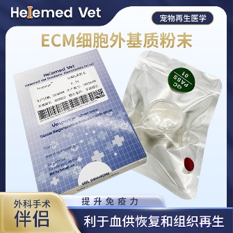
ECM Antibacterial Powder
ECM bioscaffold powder — an extracellular matrix (ECM) based antibacterial and inhibitory powder: widely used as ECM trauma dressings on infected internal/external wounds, fresh wounds, burns and crush injuries to markedly improve blood supply, accelerate healing, inhibit infection and reduce complications; it is also an ECM hemostatic material that stops capillary bleeding. How to use: after debridement, take a small amount of powder with forceps and apply to the wound surface, moisten with saline (moist wound healing) and bandage; apply once every 5 days — daily debridement is not required.

Biosynthetic Artificial Skin (6×8cm)
Artificial skin and ECM trauma dressing for external skin grafts: provides a barrier and induces regeneration, creating a good regenerative environment for subcutaneous tissue in burns and large-area skin loss.
Ask your veterinarian whether these materials suit your dog's wound; they are supplied to veterinary clinics and hospitals.
9. Clinical cases
Two cases from our partner hospitals show what chronic skin damage looks like once it has gone past "a rash": a large area of skin-muscle separation with heavy exudate, and an infected limb after orthopedic surgery with soft-tissue loss.
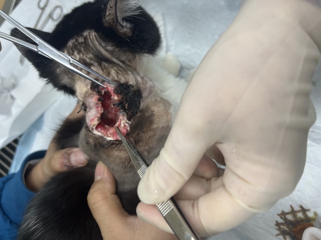
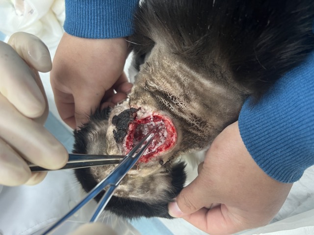
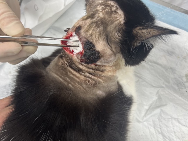
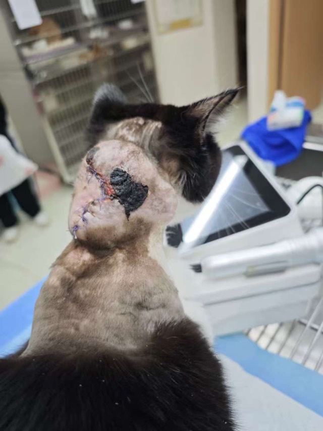
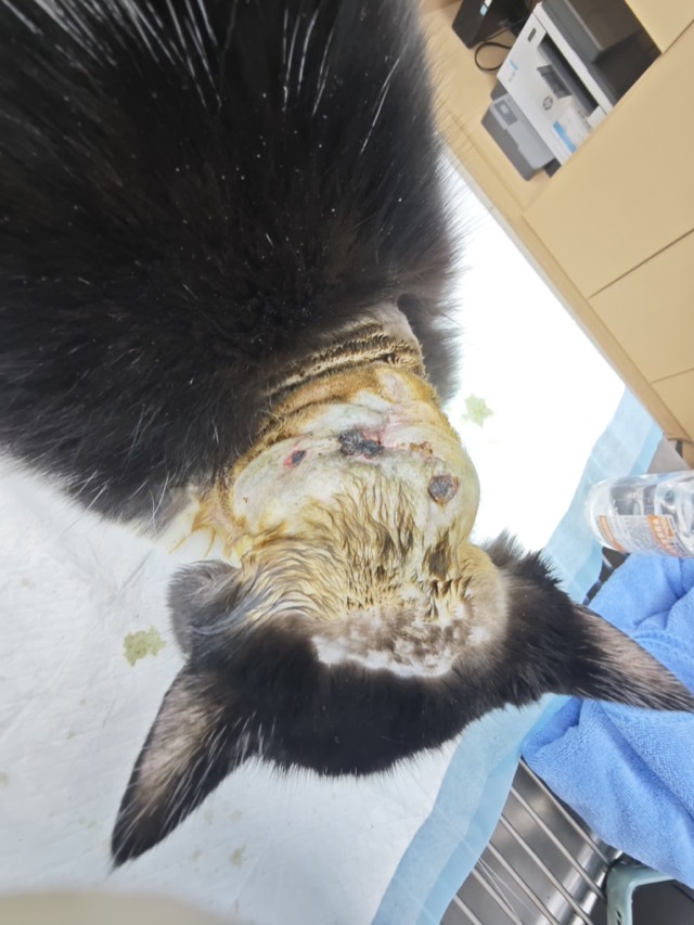
Large-Area Skin-Muscle Separation & Ulceration
A topical parasiticide burn caused large-area ulceration and heavy exudate on the neck, with skin-muscle separation and skin necrosis after the first surgery. On Feb 24 a second surgery packed two bottles of ECM Antibacterial Powder; a drain was placed the next day, the necrotic skin basically healed in 10 days, and the wound closed on Mar 4.
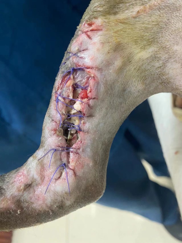
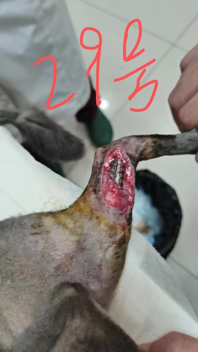
Orthopedic Infection with Soft-Tissue Loss
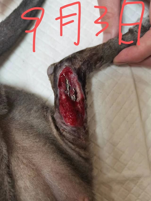
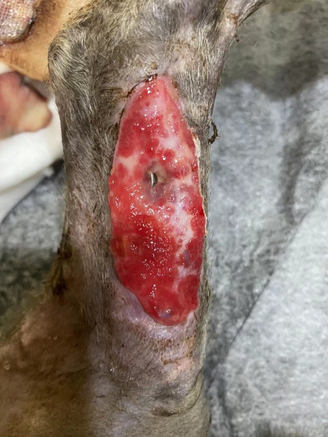
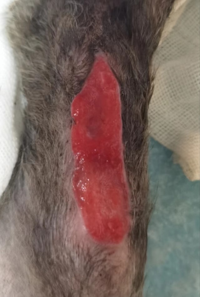
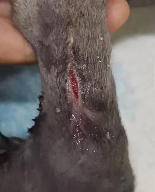
Open infected fracture with post-op infection, soft-tissue loss and severely exposed bone plate; after debridement ECM powder was placed on the wound area, moistened with saline and bandaged. Granulation began on day 3, grew markedly by day 5 and gradually enveloped the plate, skin started growing at the 6th application, and it ultimately basically recovered.
Cases from partner veterinary hospitals. ECM materials are veterinary surgical materials and are applied by the treating veterinarian. See all 39 clinical cases →
Frequently asked questions
How do I know if my dog has a bacterial or a yeast skin infection?
You cannot reliably tell by looking. Greasiness, thickening, dark pigmentation and a musty smell point towards yeast; pustules, circular scaling lesions and crusts point towards bacteria; most chronic cases have both. A stained smear examined under the microscope takes minutes and answers it properly.
Is a dog skin infection contagious to humans or other pets?
Bacterial and yeast infections are not usually passed to people, though resistant staphylococci can occasionally spread. Ringworm and sarcoptic mange are genuinely contagious to people and other pets, which is why a vet should look at any new skin lesion rather than starting an over-the-counter shampoo.
How long does it take to treat a dog skin infection?
Topical therapy usually runs for three to four weeks. If oral antibiotics are needed, three to four weeks is the minimum, continued for one to two weeks beyond clinical cure. Stopping when the coat looks normal is the most common reason the infection returns.
Why does my dog keep getting skin infections?
Almost always because of an underlying disease: environmental or flea allergy is the most common, followed by food allergy, hypothyroidism and Cushing’s disease. Repeated short antibiotic courses treat the symptom and leave the cause untouched. Ask your vet to work up the underlying disease.
Can I treat my dog’s skin infection at home?
Medicated shampoos and sprays prescribed by your vet are the mainstay of home treatment, and they matter a lot. What you should not do is use human antibiotic or steroid creams, human antifungal preparations, or leftover tablets from a previous course.
What does a deep skin infection look like?
Nodules, draining tracts, ulcers, marked swelling and pain, sometimes with fever and loss of appetite. Deep pyoderma needs bacterial culture and a longer course of treatment than a surface infection, and it should never be managed with shampoo alone.
Will a skin infection heal by itself?
Superficial infections sometimes improve temporarily, but without addressing the cause the dog re-traumatises the skin and the infection returns. Deep infections do not resolve on their own and can spread. Treating early is both kinder and cheaper than treating a chronic case.