
Case Gallery
Real cases from front-line animal hospitals across China — excerpted from “Amazing ECM Clinical Case Sharing” (106 pages), with 39 cases and 29 clinical surgical videos. Click an image to enlarge and view details.
Soft-Tissue Wound Regeneration · ECM Antibacterial Powder / ECM Sponge
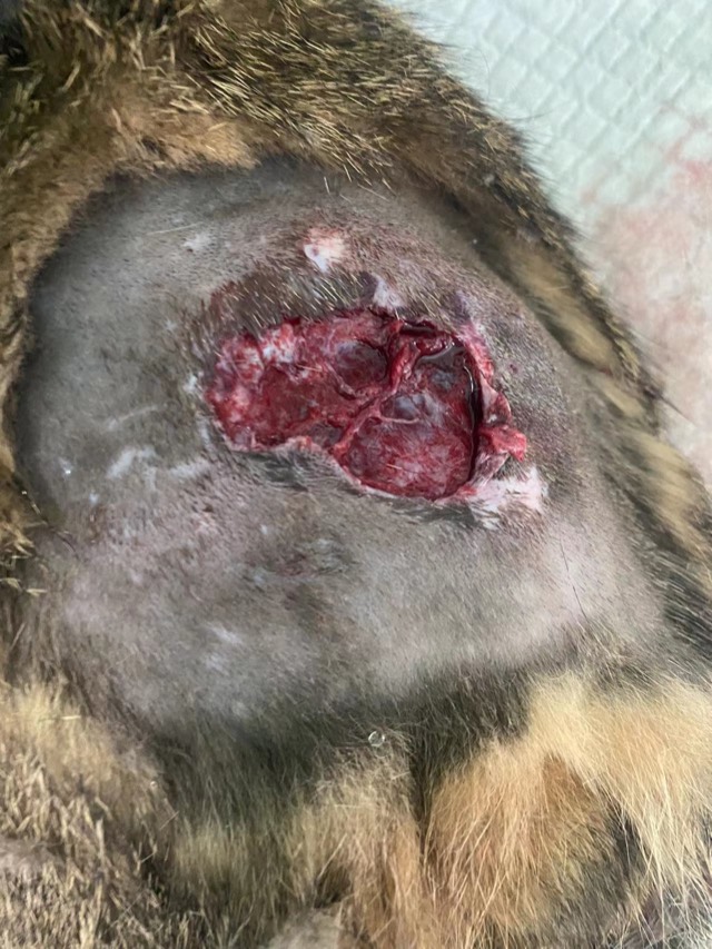
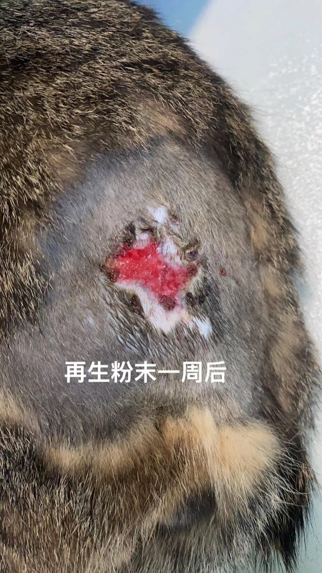
Bite-Wound Regeneration
After debridement and irrigation of the bitten, ulcerated wound, ECM Antibacterial Powder (ECM Inhibitory Powder) was placed on the wound, moistened with saline and bandaged with gauze. Re-check one week later showed the wound markedly reduced, with normal soft-tissue and surrounding-skin regeneration, no infection and no premature scabbing.
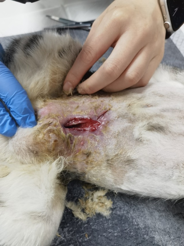
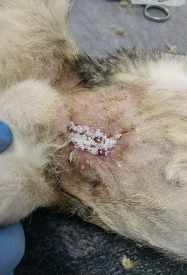
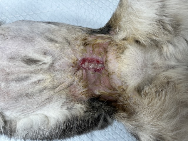
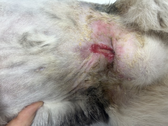
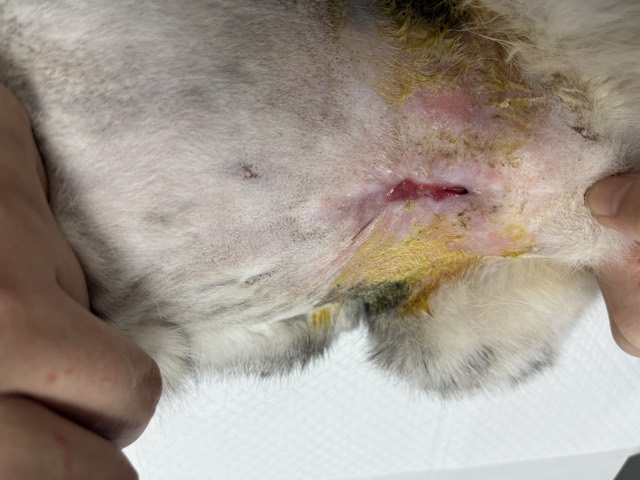
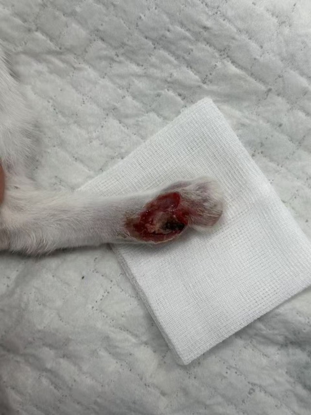
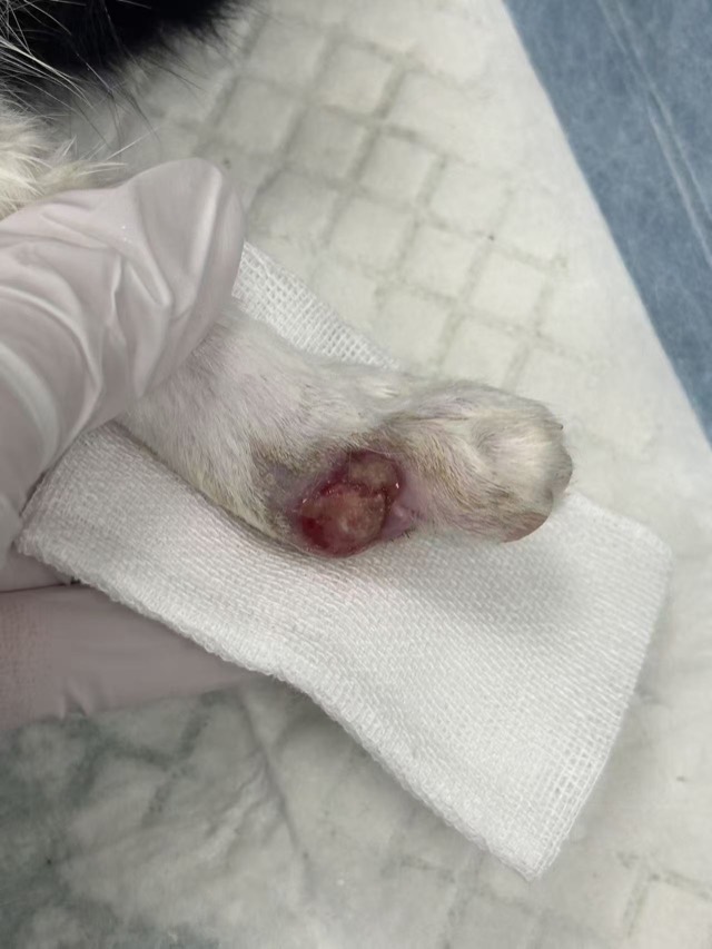
Small-Cavity Wound Healing
The wound failed to heal after more than a month of conventional treatment. ECM Antibacterial Powder was applied and bandaged on Jul 23; after two application cycles (10 days) the wound healed markedly with no infection or premature scabbing, and it fully recovered after one more cycle.
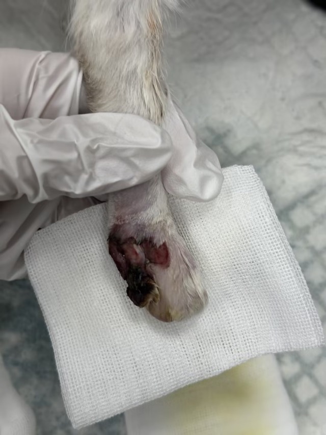
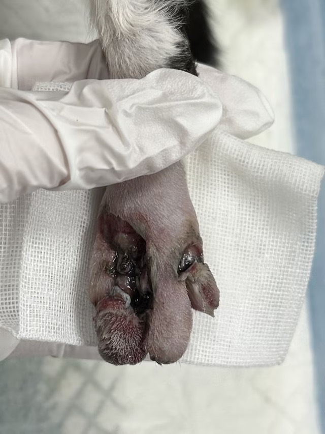
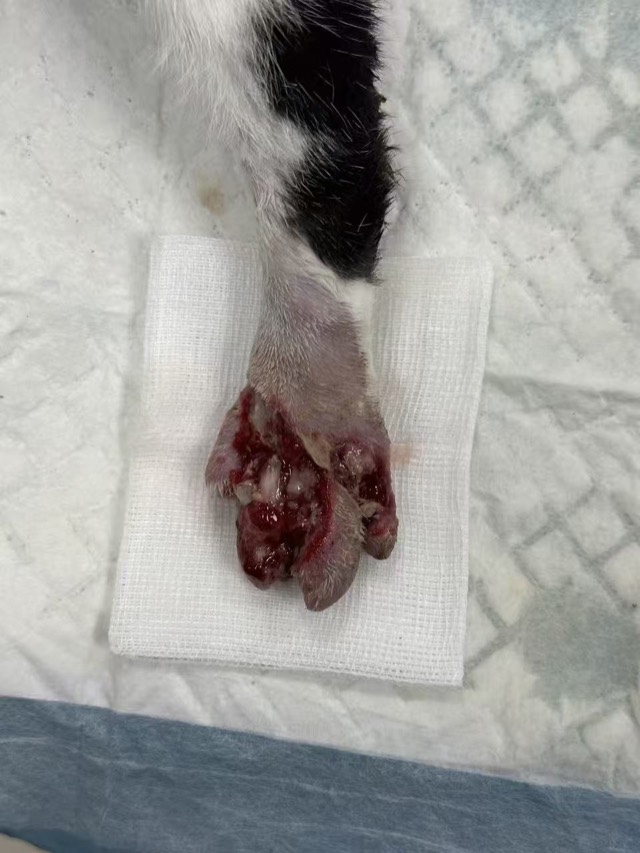
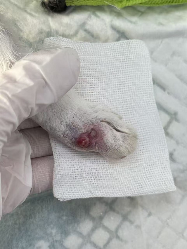
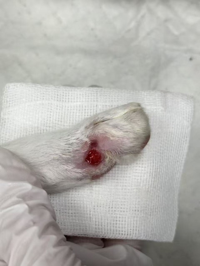
Kitten Toe Elevator Crush Injury
A two-month-old stray kitten had both feet caught in an escalator, with skin-muscle separation and necrosis. Half a bottle of ECM Antibacterial Powder was packed in initially, moistened with saline and bandaged, then dressed cyclically: infection controlled, blood supply restored, granulation grew markedly, and the wound basically recovered in about one month.
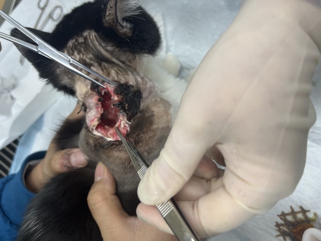
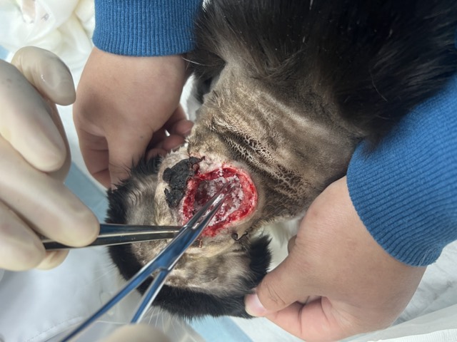
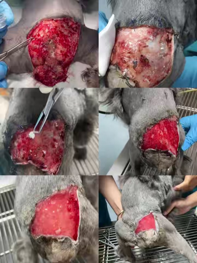
Large-Area Skin-Muscle Separation & Ulceration
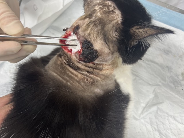
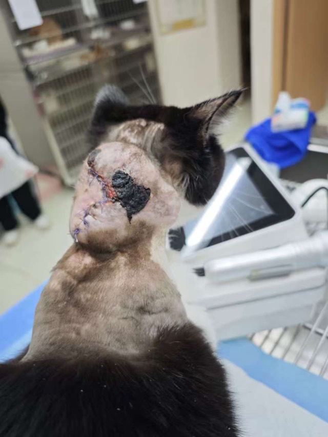
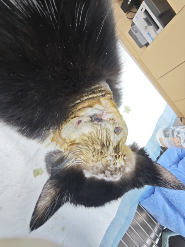
A topical parasiticide burn caused large-area ulceration and heavy exudate on the neck, with skin-muscle separation and skin necrosis after the first surgery. On Feb 24 a second surgery packed two bottles of ECM Antibacterial Powder; a drain was placed the next day, the necrotic skin basically healed in 10 days, and the wound closed on Mar 4.
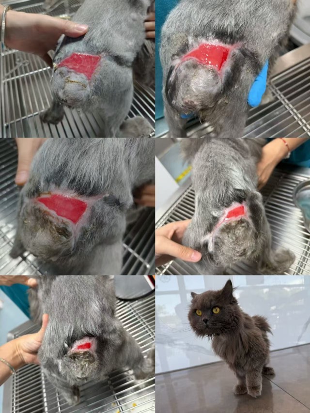
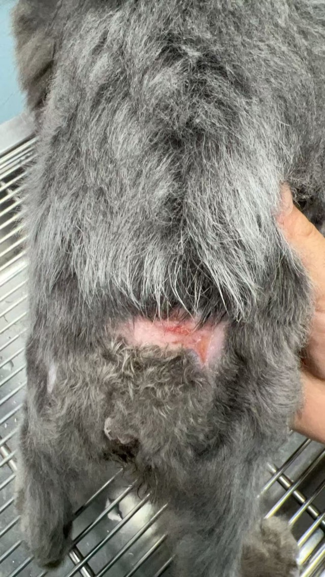
Tail Fracture and Hind-Limb Necrosis (Cola)
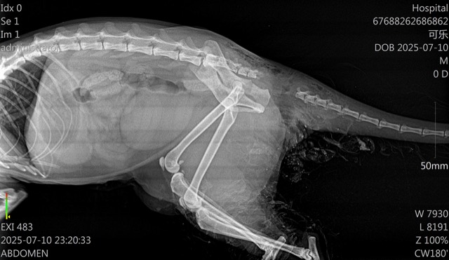
At transfer the fracture site felt fluid-like and emitted a foul odor; debridement revealed rot from the sacrum to the tailbone and left hind-limb suppuration up to above the calcaneus. After four irrigations and thorough debridement, two boxes of ECM Antibacterial Powder were applied and wrapped in sterile gauze. The wound kept shrinking with staged dressing changes; it basically healed on Aug 26, admitted Jul 12 and discharged recovered on Aug 31 — one month in total.
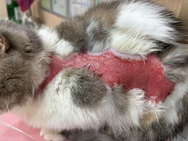
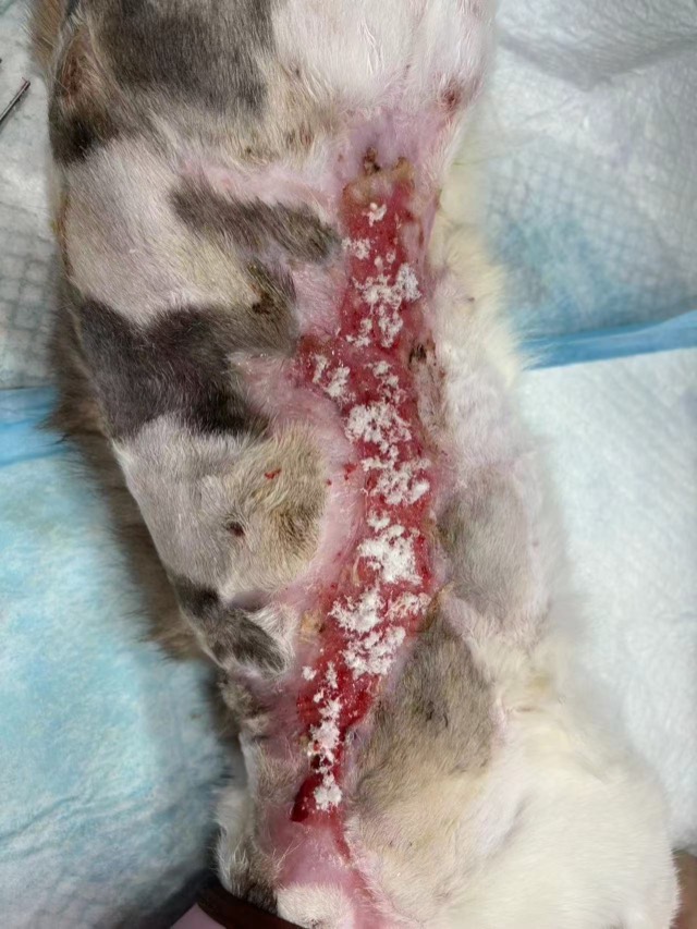
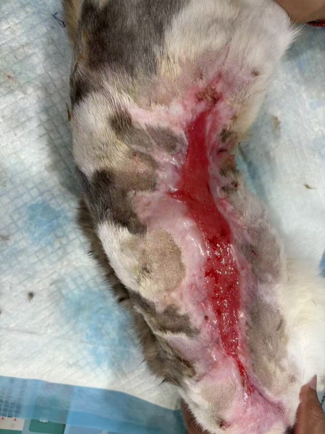
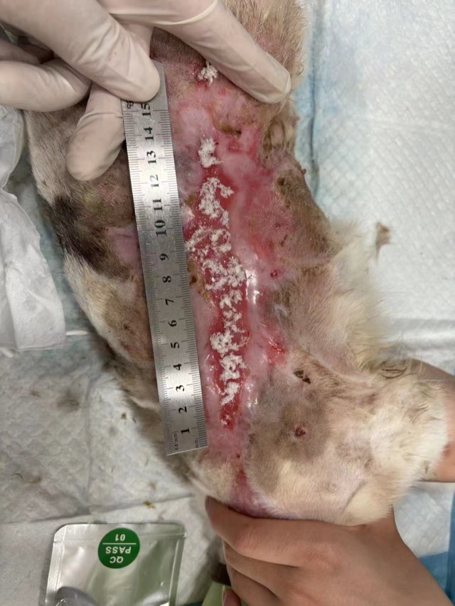
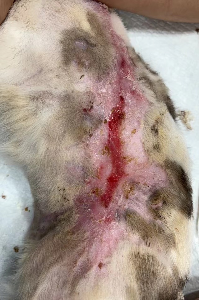
Large-Area Burn (approx. 40cm)
An adult cat with an approx. 40cm large-area burn on its side. ECM Antibacterial Powder was first applied on Sep 11, 2024; after 5 days the wound was markedly reduced with no infection. Areas hard to bandage were fixed with skin staples or a stitch at each corner. The wound kept shrinking with each application and gradually recovered after Oct 8.
Car-Crash Crush Wound
A car-crash crush caused large-area soft-tissue damage; after debridement ECM Antibacterial Powder was applied to promote blood-supply recovery and soft-tissue regeneration.
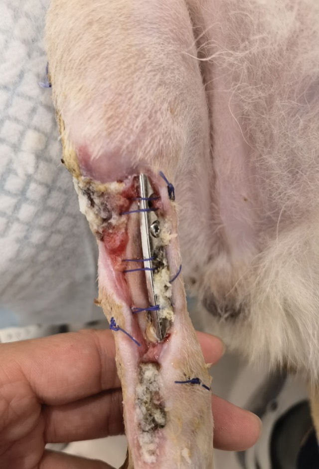
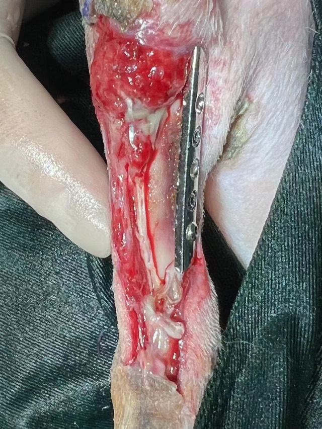
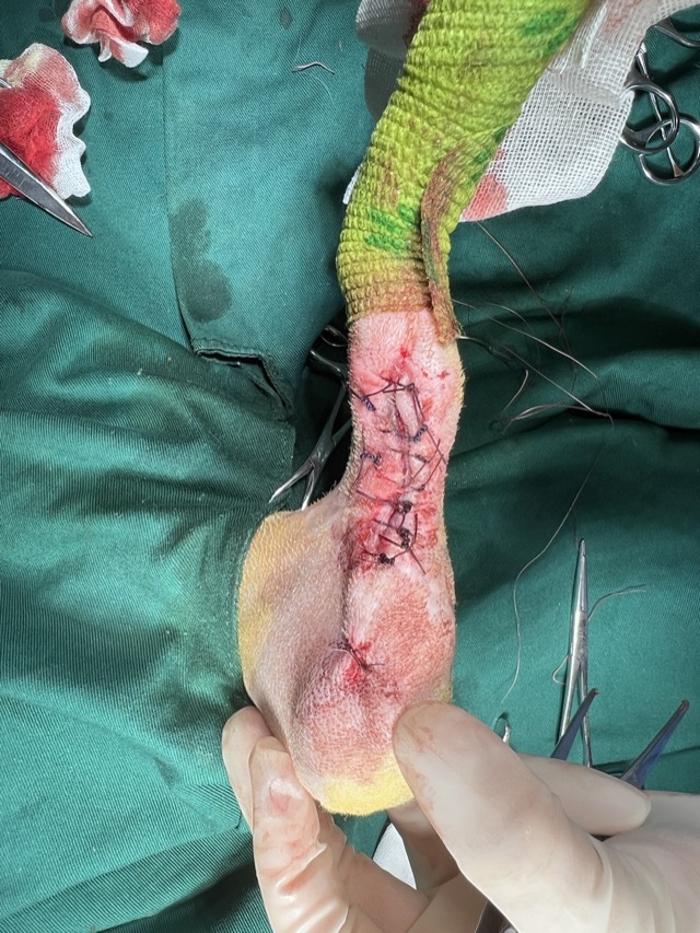
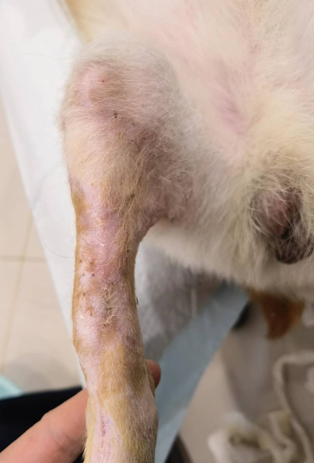
Post-Orthopedic-Surgery Infection
Post-orthopedic-surgery infection treated with ECM Antibacterial Powder; after 3 application cycles (15 days) the bone plate was removed and a skin flap sutured, and the wound fully recovered.
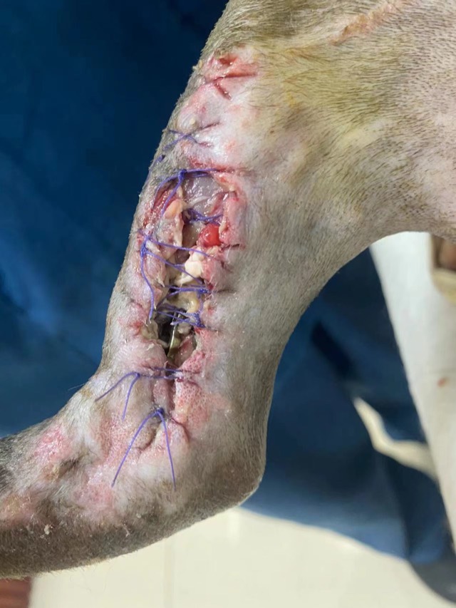
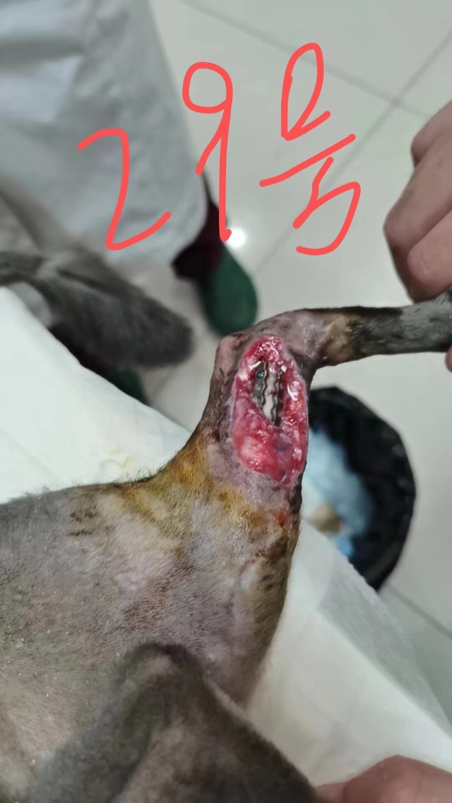
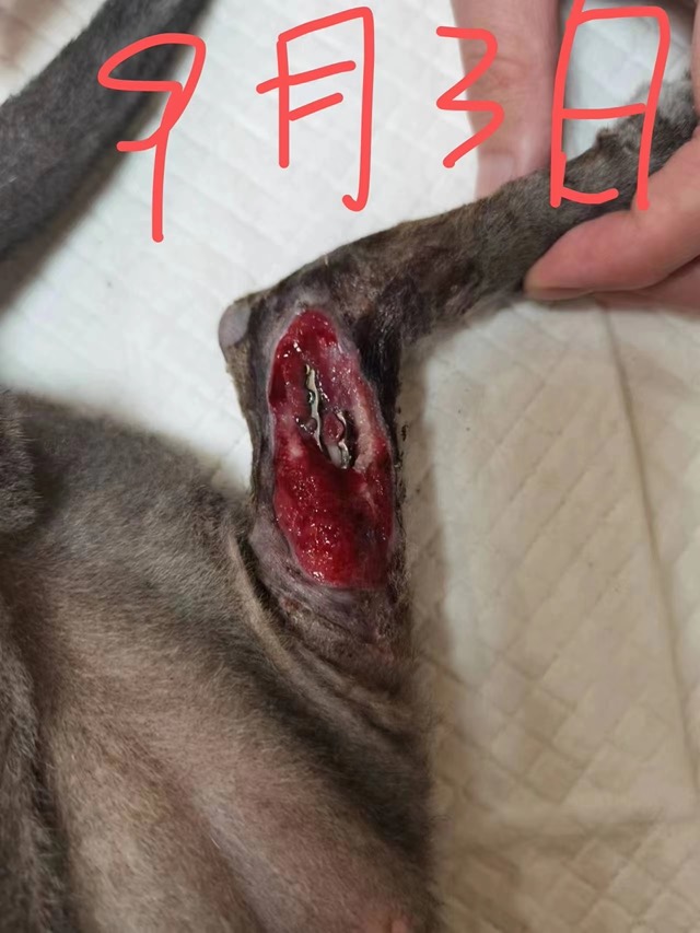
Orthopedic Infection with Soft-Tissue Loss
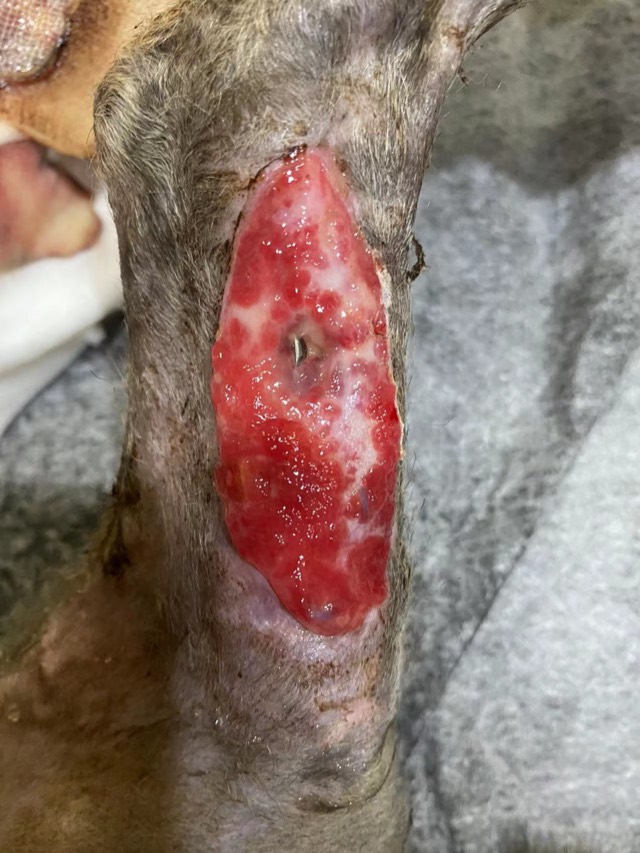
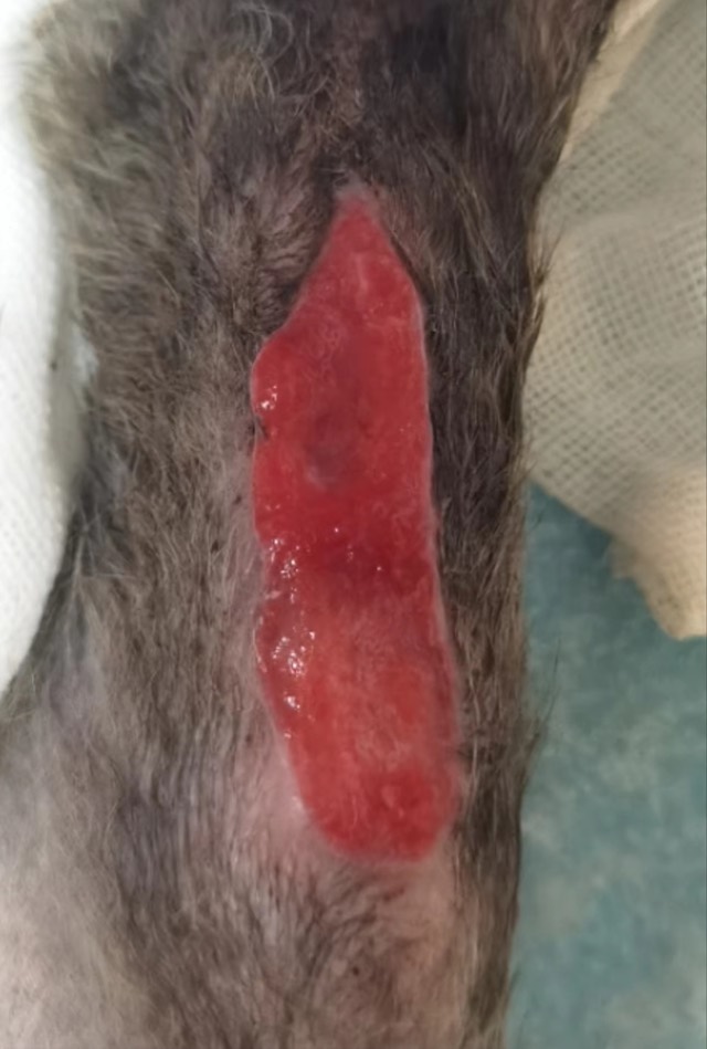
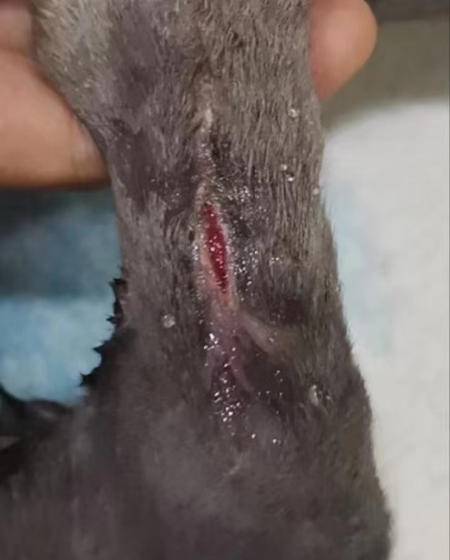
Open infected fracture with post-op infection, soft-tissue loss and severely exposed bone plate; after debridement ECM powder was placed on the wound area, moistened with saline and bandaged. Granulation began on day 3, grew markedly by day 5 and gradually enveloped the plate, skin started growing at the 6th application, and it ultimately basically recovered.
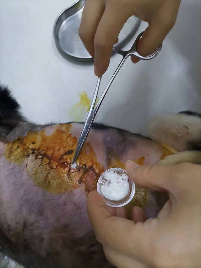
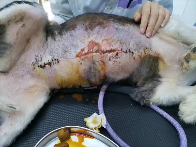
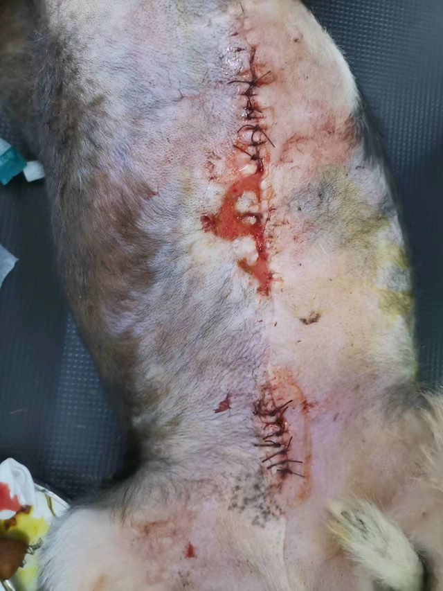
Non-Healing Wound after Mastectomy
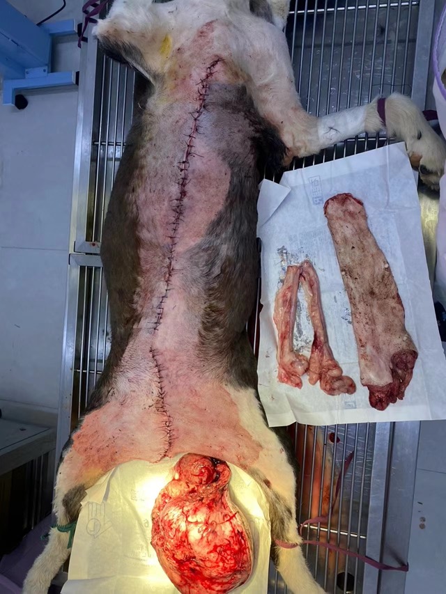
Thirty days after mastectomy the chest wound would not heal; ECM powder was applied subcutaneously and on the surface then bandaged. After 5 days clear signs of regeneration appeared, and it basically recovered after two applications (10 days).
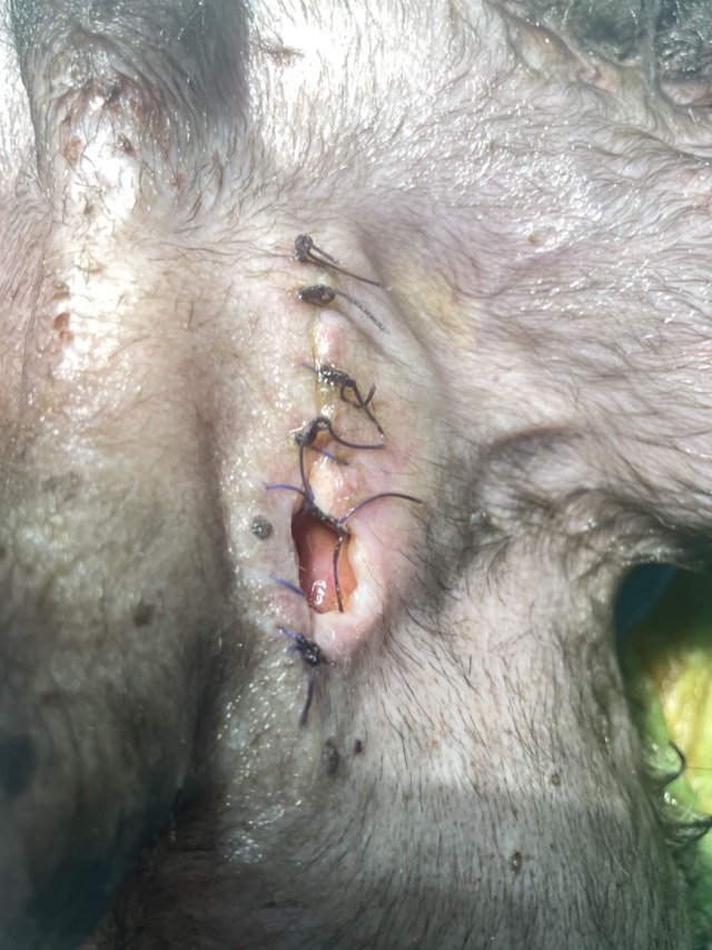
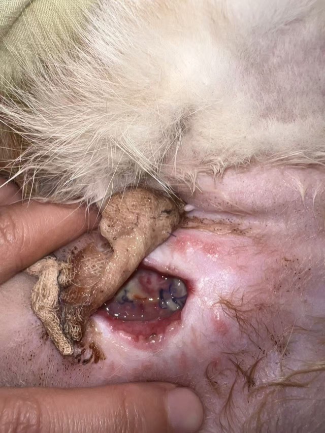
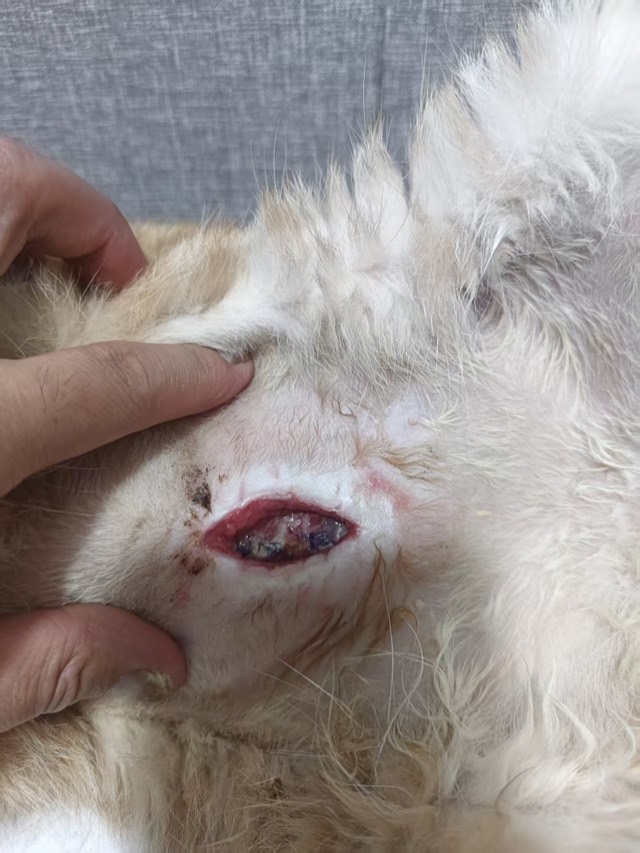
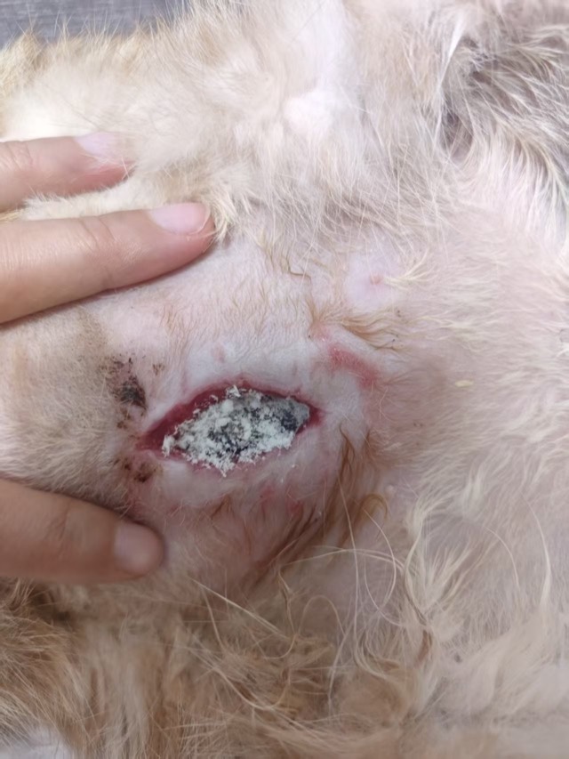
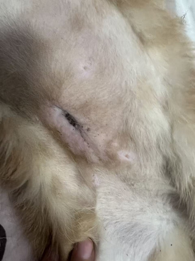
Fat Liquefaction after Bladder-Stone Surgery
After bladder-stone surgery subcutaneous fat liquefaction and non-healing wound; after thorough debridement an appropriate amount of ECM powder was placed on the subcutaneous wound surface and the wound sutured in a single application. First powder on Aug 29, second on Sep 1, and the wound consolidated on Sep 11.
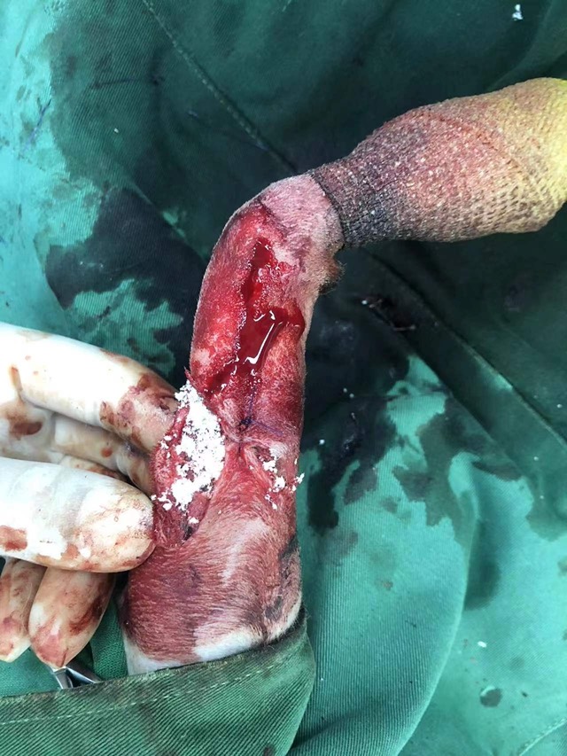
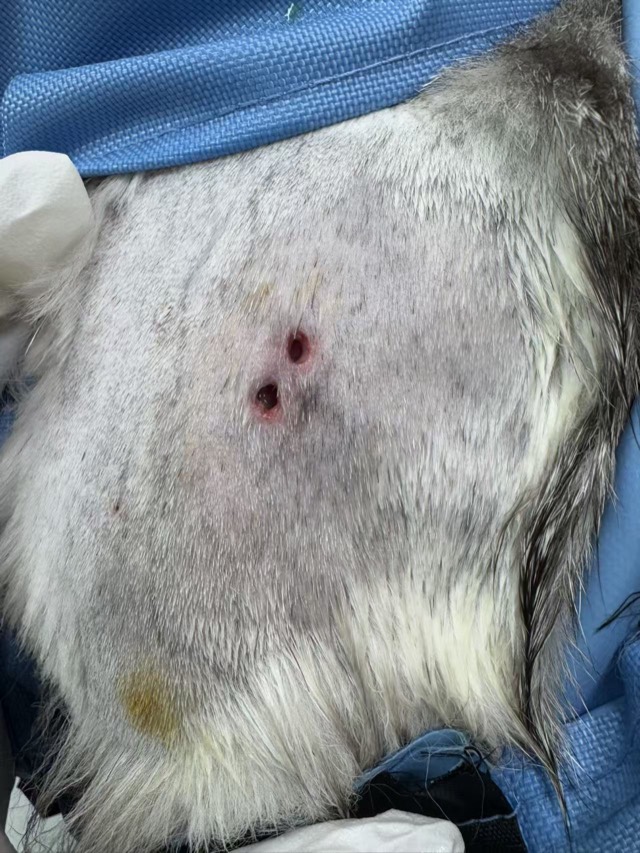
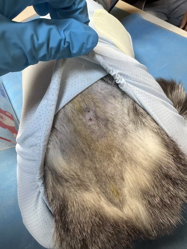
Prophylactic Bite-Wound Application (Subcutaneous Injection)
Bite cases often develop large subcutaneous skin-muscle separation; injecting the ECM Antibacterial Powder–saline suspension subcutaneously in multiple sessions via syringe markedly speeds primary recovery and avoids accidents. Applied May 1, the separated area recovered by May 10.
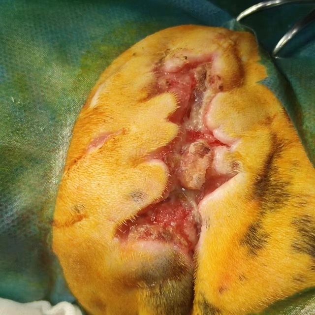
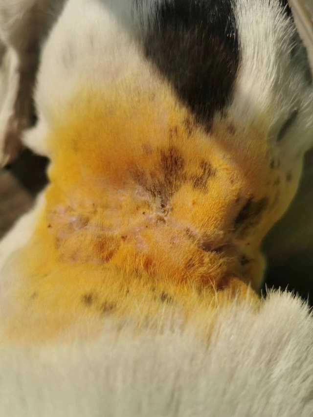
Transferred Bite-Wound Case
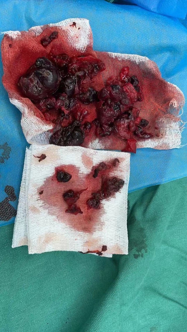
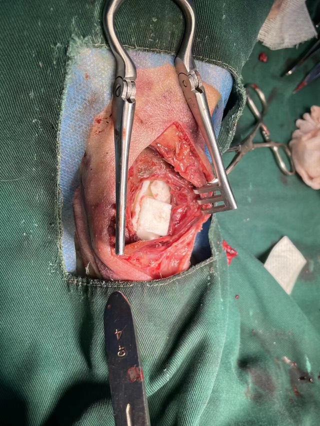
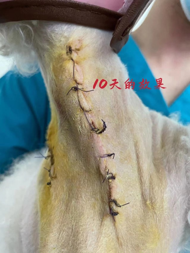
Treated at another hospital for about 20 days (surgical debridement + antibiotics); at transfer the wound was non-healing with large amounts of purulent infected tissue underneath. After sedation the subcutaneous wound was thoroughly debrided and a thin layer of ECM powder sprinkled on the surface and bandaged.
ECM Sponge Assisted Cavity Filling
When there is an obvious cavity with normal surrounding blood supply, ECM Sponge can assist filling (larger volume, ~20-day degradation, synchronized with new tissue). Before use, fully soak in saline and squeeze out excess water.
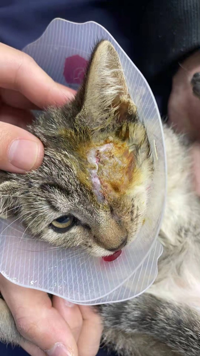
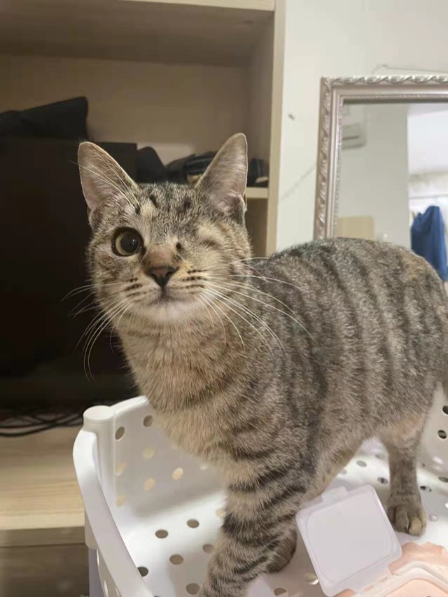

Enucleation Cavity Filling
After enucleation, ECM powder and ECM Sponge were placed to reduce exudate and speed recovery; at 2 months it remained full — the sponge had been replaced by autologous new tissue. Without filling there would inevitably be a depression and a permanent subcutaneous cavity. Apply moderately; more is not better.
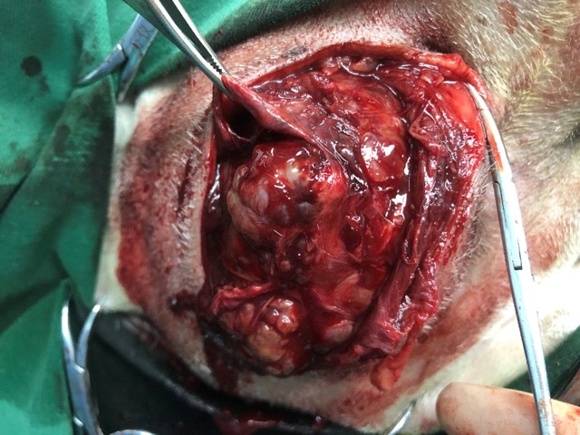
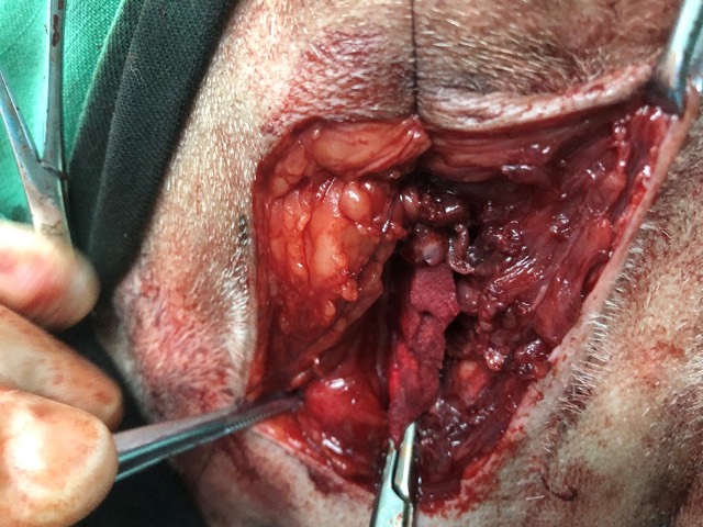
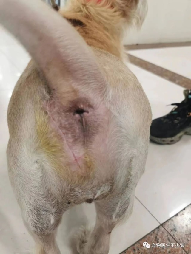
Tumor-Resection Cavity Filling
Tumor-resection surgery often has poor post-op healing; where an obvious cavity exists, implant ECM Sponge for filling (like tissue grafting), lay ECM powder subcutaneously for prophylaxis, then close the wound — effectively accelerating primary healing. Two pieces of ECM Sponge were implanted in this case.
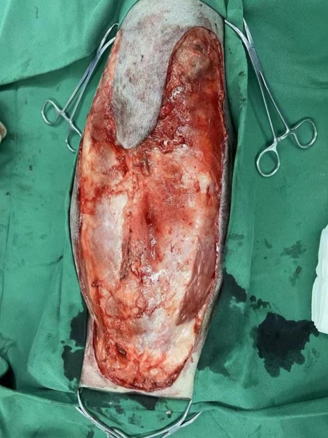
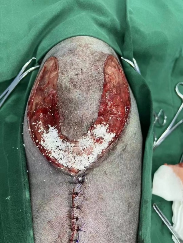
Prophylactic Total Mastectomy Application
The complication rate of total mastectomy is second only to bite cases, so prophylactic application is especially recommended: lay a thin layer of ECM powder subcutaneously in the triangular, high-tension area; use ECM Sponge prophylactically in the inguinal region to avoid cavities and accelerate tissue regeneration.
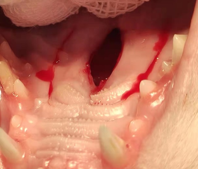
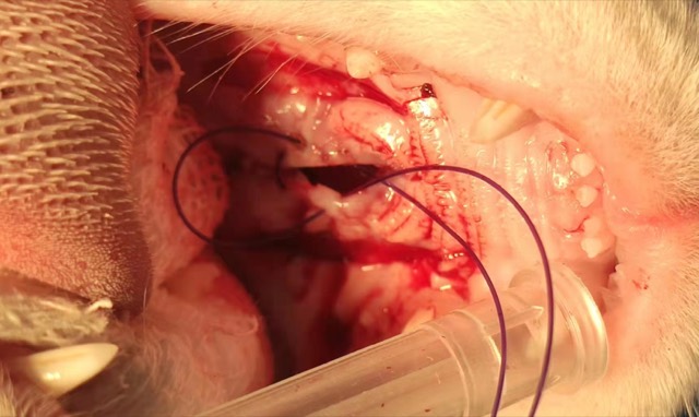
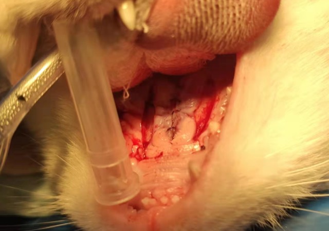
Cleft Palate Repair and Regeneration
After creating a fresh wound on the cavity wall below the cleft palate, ECM Sponge was packed; after adequate tension release it was sutured, using mid-term monofilament absorbable suture with the knot placed subcutaneously to reduce irritation.
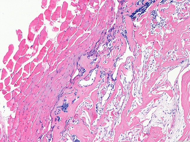
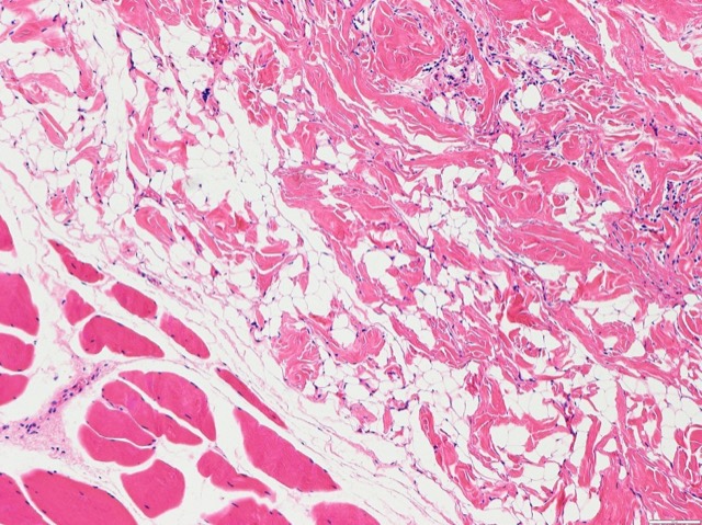
ECM Sponge In-Vivo Fate Follow-up
X-ray follow-up of the ECM Sponge's in-vivo fate after implantation: the sponge gradually degrades and is replaced by autologous new tissue (2–26 weeks), achieving true tissue repair rather than a permanent foreign body.
Prophylactic Orthopedic Application (Subcutaneous Powder)
Before suturing the skin flap in orthopedic surgery, placing an appropriate amount of ECM powder subcutaneously accelerates vascularization and soft-tissue healing, benefits bone regeneration, and avoids exudate and infection. Timely prophylactic application is recommended for comminuted and open fractures to speed primary healing.
Standard ECM Antibacterial Powder Application Method
① After thorough debridement, evenly sprinkle a thin layer of ECM powder on the wound surface; ② moisten with saline; ③ gauze bandage to keep moist; ④ wrap with a bandage to secure; ⑤ powder change cycle is 5 days, no daily dressing change needed in between.
Bone Tissue Regeneration · Antibacterial Artificial Bone / Artificial Bone Plus (ECM Bioscaffold)
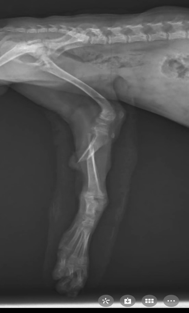
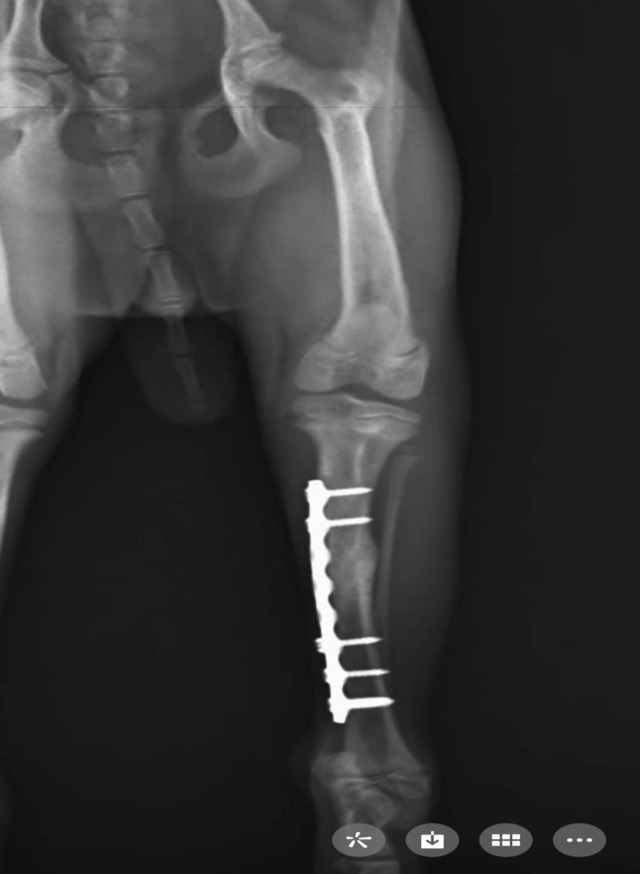
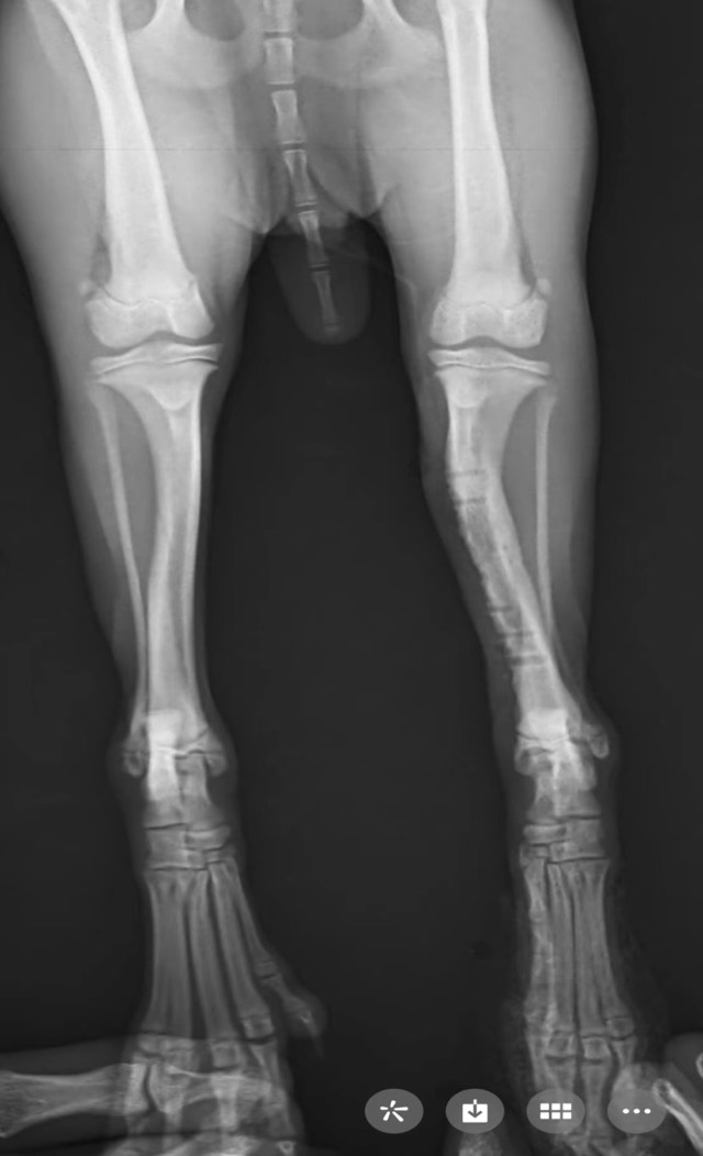
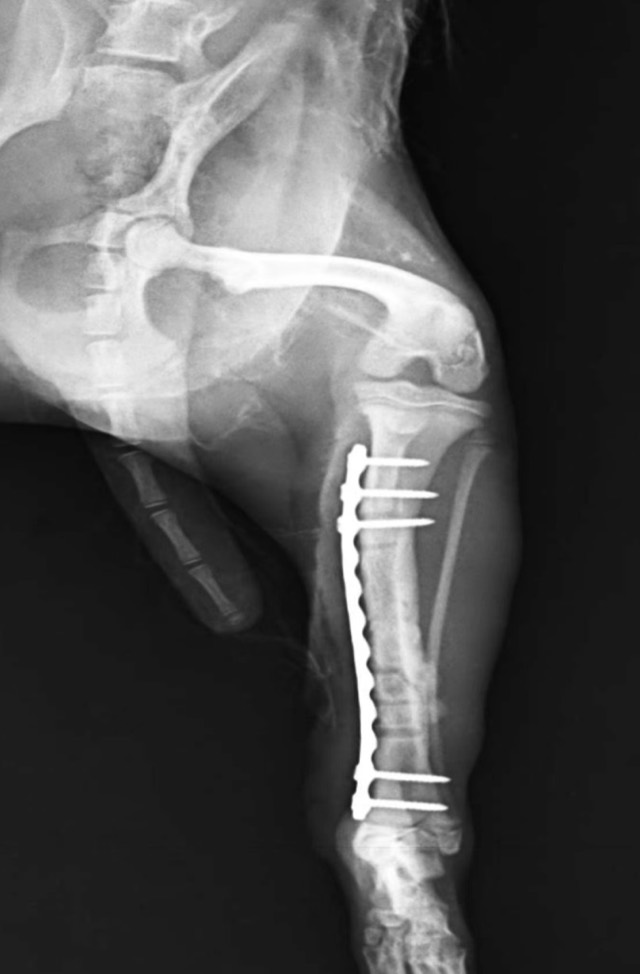
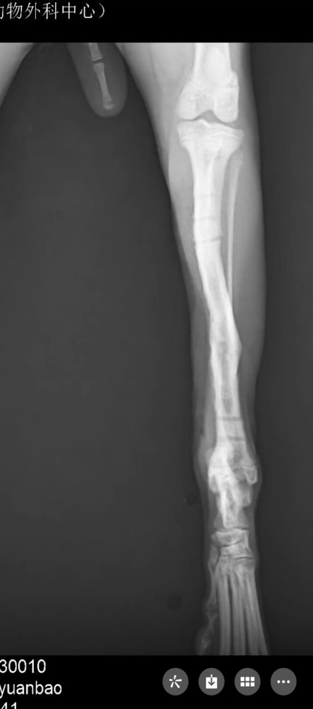
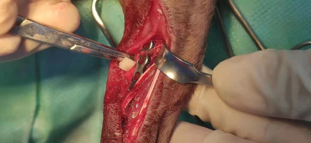
Tibial Oblique Fracture Re-Fracture Revision
After the first internal fixation of a tibial oblique fracture and plate removal following normal recovery, a secondary fracture appeared at the third screw hole one week later (common in small animals). The second fixation used Antibacterial Artificial Bone granules — filling the defect where present, or around the fracture line where not; the new bone density was markedly higher. At plate removal after healing, Antibacterial Artificial Bone was packed into the screw holes to accelerate osteogenesis, and no further secondary fracture occurred.
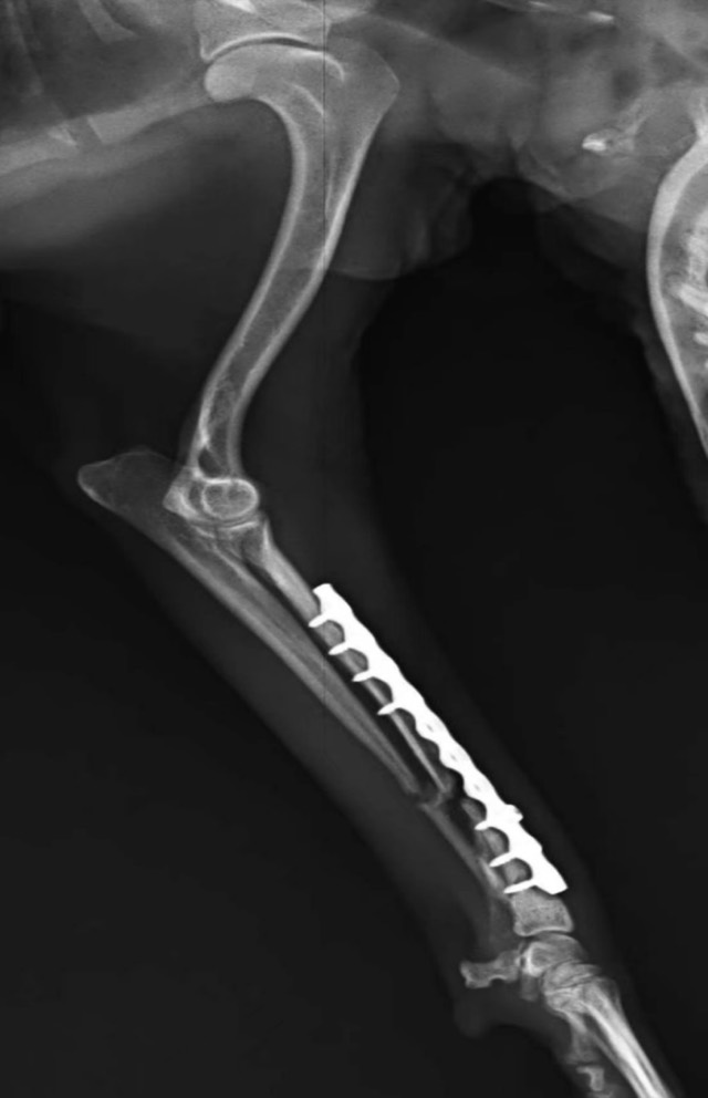
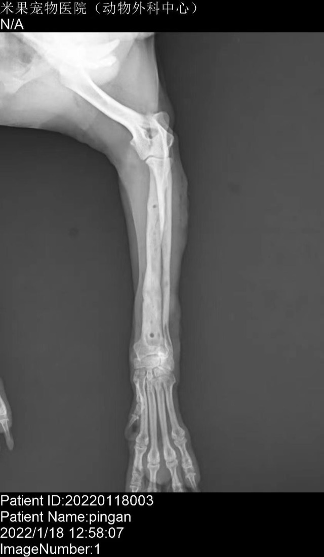
Post-Op Radial/Ulnar Bone Dissolution (Case 1)
Bone dissolution appeared at the 30-day re-check after the first surgery; the site was opened and Antibacterial Artificial Bone applied to raise local calcium and phosphorus concentration. After all plates were removed, bone healing was good with no secondary fracture.
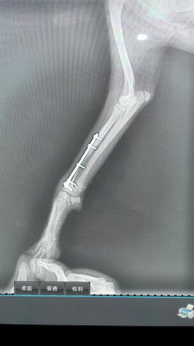
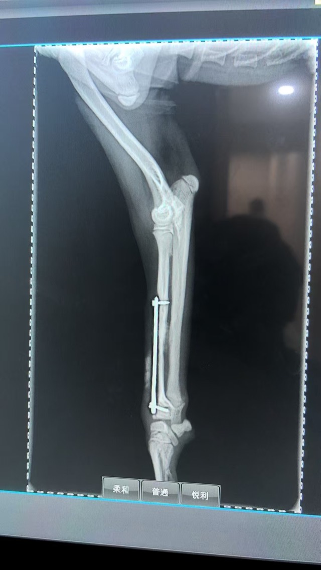
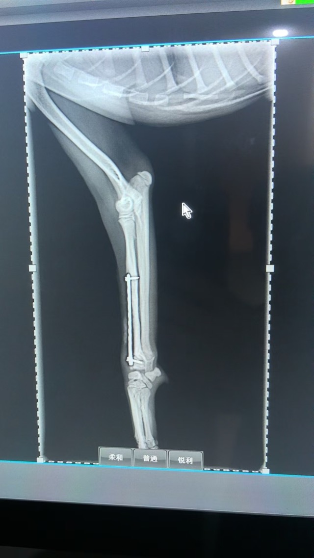
Post-Op Radial/Ulnar Bone Dissolution (Case 2)
The owner missed the plate-removal window and only returned after two to three months, with bone dissolution. The middle screw was removed and artificial bone powder packed in; growth was rapid with strong activity.
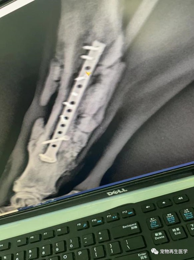
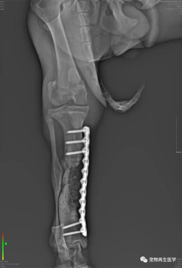
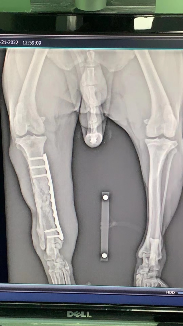
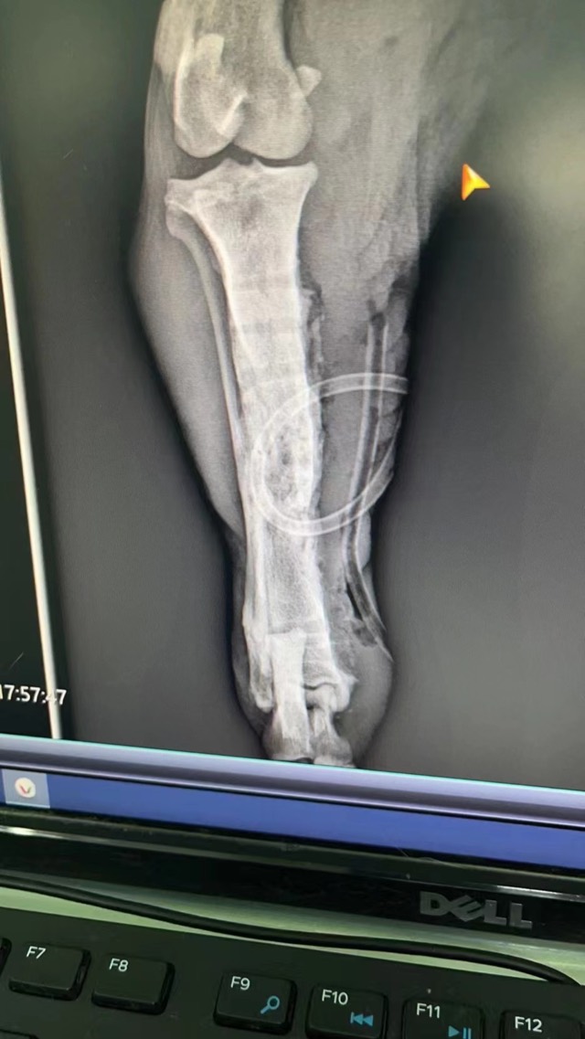
Old Comminuted Fracture Revision
A third revision surgery for severe comminuted, infected fracture with very large bone defect; Artificial Bone Plus (ECM bioscaffold with added BMP) was used together with a periosteal patch — isolating soft tissue to prevent premature invasion and forming early periosteum through vascularization to nourish osteogenesis. Eventually the plate was removed and movement recovered as before.
Fracture Fixation and Bone Grafting (X-ray Follow-up)
Bio-artificial bone (ECM + bioactive ceramics) was used for fracture fixation and bone grafting; X-ray follow-up showed new bone formation and increased bone density.
Dental Regeneration · Alveolar Bone Reconstruction / Periodontal GTR Guided Regeneration
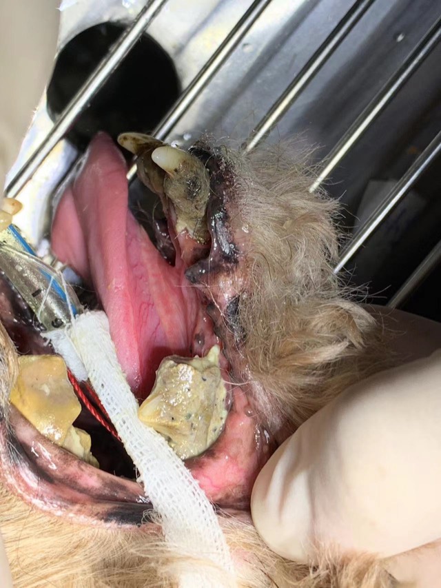
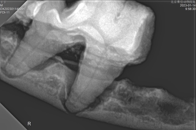
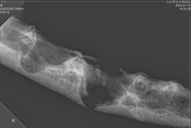
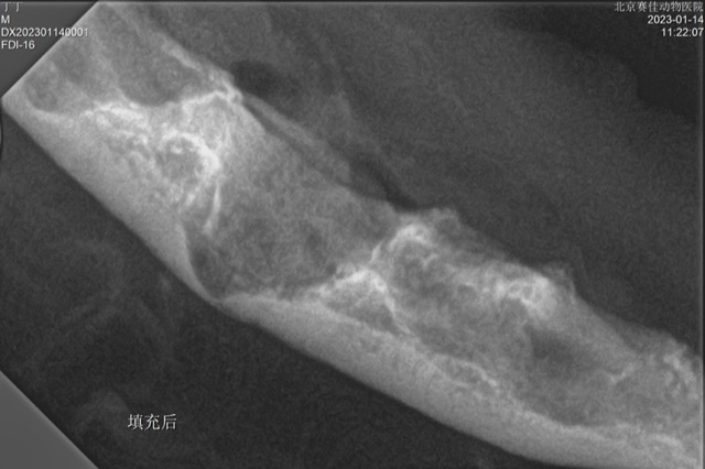
Tooth Extraction and Alveolar Bone Reconstruction
After extraction, site preservation surgery is needed: alveolar bone regeneration and alveolar bone reconstruction prevent fracture, oronasal fistula, facial fistula and other risks. X-ray showed mandibular dissolution and defect; after filling with Antibacterial Artificial Bone granules (alveolar bone filling material) the alveolar bone was regenerated. No infection or fracture post-op, and re-check showed good mandibular regeneration.
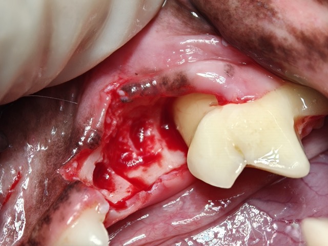
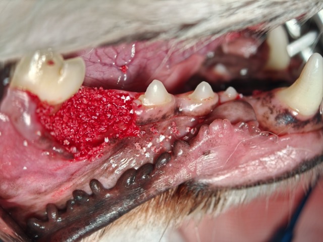
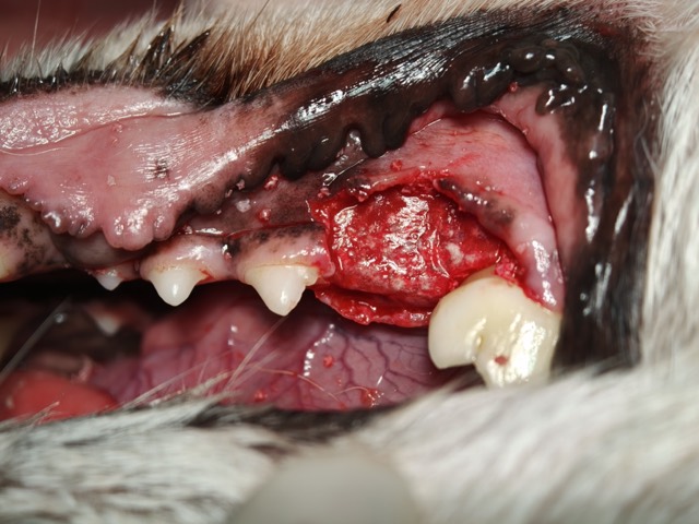
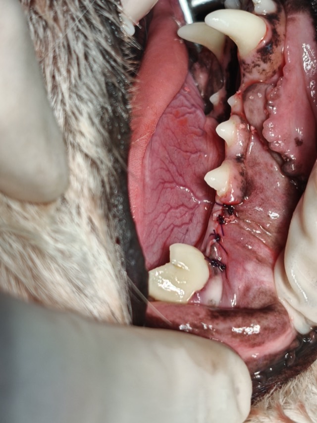


Standard Four Steps of Alveolar Bone Reconstruction
Standard workflow for extraction and alveolar bone reconstruction (site preservation surgery): ① extract and curette → ② fill Antibacterial Artificial Bone → ③ cover with Regeneration Barrier Membrane (isolating soft tissue) → ④ suture gingiva. Antibacterial Artificial Bone fills the socket, the membrane covers and isolates it from soft tissue, then gingiva is sutured — the same sequence used for subgingival regeneration and periodontal tissue regeneration; re-check at 3 months showed perfect, complete mandibular reconstruction.
Periodontal GTR Guided Regeneration (Tooth-Saving)
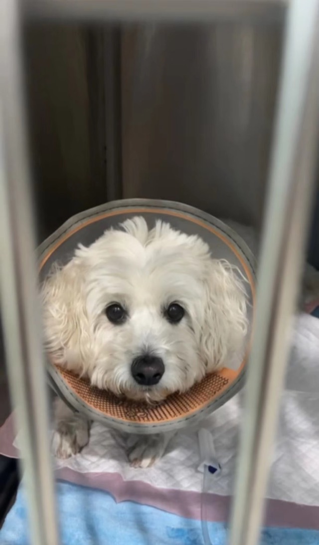
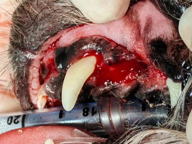
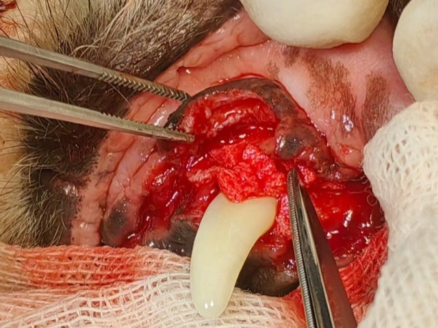
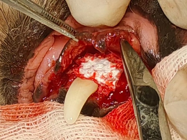
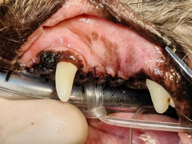
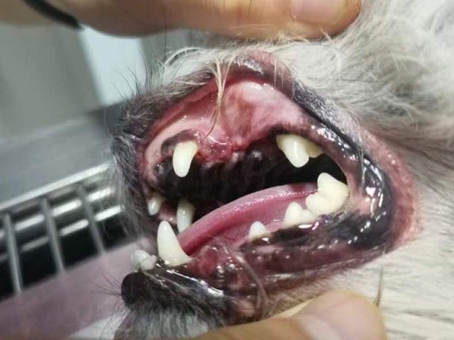
A 10-year-old dog with severe periodontal disease — swollen and inflamed gums and alveolar bone atrophy; the owner declined multiple extractions and chose GTR tooth preservation (GTR tooth-saving surgery): mucosal flap reflection, extraction of the periapical-periodontitis tooth, root planing and repeated curettage, Antibacterial Artificial Bone granules packed and covered with Regeneration Barrier Membrane (smooth side inward), tension-free nodular suturing, and “button” principle to tighten the periodontal space around the canine. The goal of the procedure was periodontal regeneration, subgingival regeneration and periodontal tissue regeneration at the same site. At 10-day suture removal tooth stability was better, with good follow-up.

Oronasal Fistula Filling and Hemostasis
Oronasal fistula occurs not only in unextracted cases but also commonly after extraction — improper suturing leaves a hidden risk, and the same defect can open into a facial fistula. If the cavity is large and bleeding is heavy during mucosal flap reflection, cut ECM Sponge to size and pack the fistula tract, then do a flap suture, hooking the sponge with a few stitches to prevent inhalation into the nasal cavity. The sponge fully grows into soft tissue and blocks the fistula.
Hernia & Chest-Wall Repair · Biosynthetic Absorbable Hernia Mesh
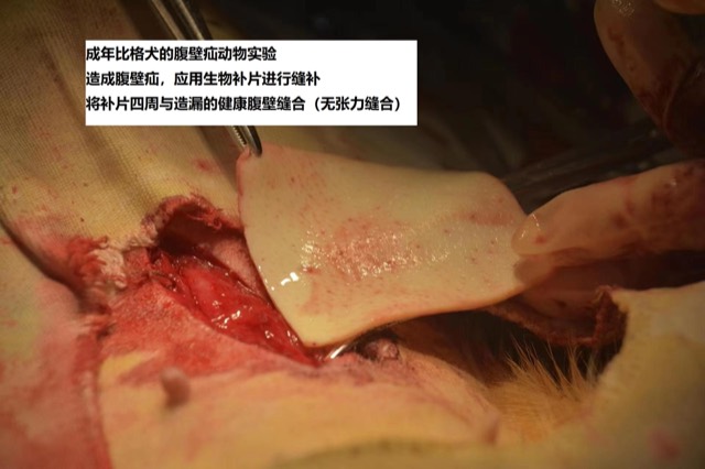
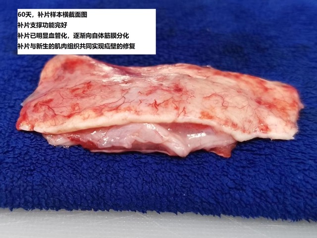
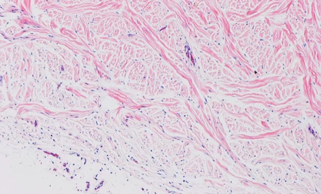
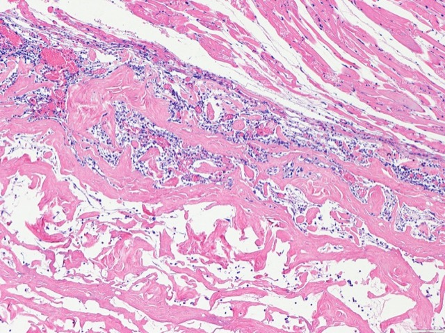
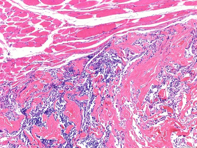
Hernia Mesh Animal Experiment & Pathological Validation
Biosynthetic Absorbable Hernia Mesh animal experiment: after implantation, cells and vessels grew in, fully rebuilding the hernia region; the 60-day pathological section showed fibroblasts infiltrating the mesh with abundant differentiated mature fibrocytes and vascular distribution, and no inflammatory reaction.
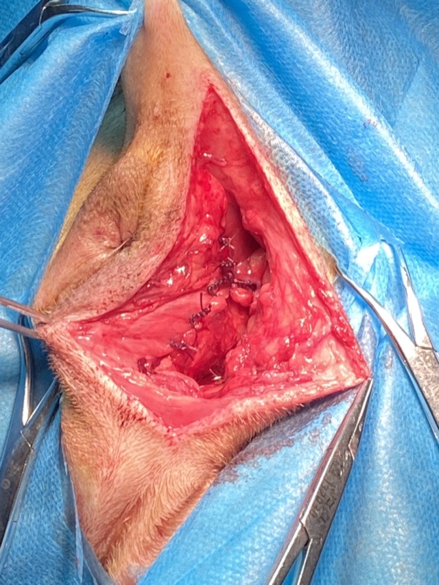
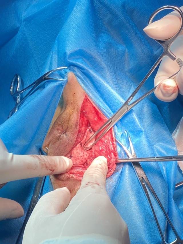
Perineal Hernia Repair
The only domestic biosynthetic hernia mesh (porcine decellularized process): first close or minimize the hernia ring, then suture the mesh to the surrounding healthy tissue; its natural pores make needle entry easy without destroying physiological structure. Early support shares tension; later cells and vessels grow in for full reconstruction — biodegradable with no long-term foreign-body sensation.
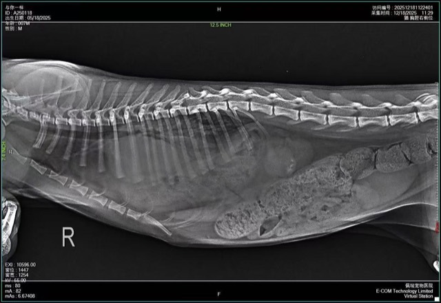
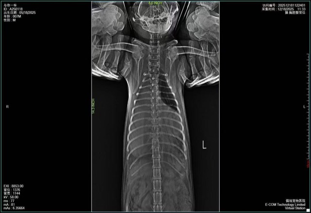
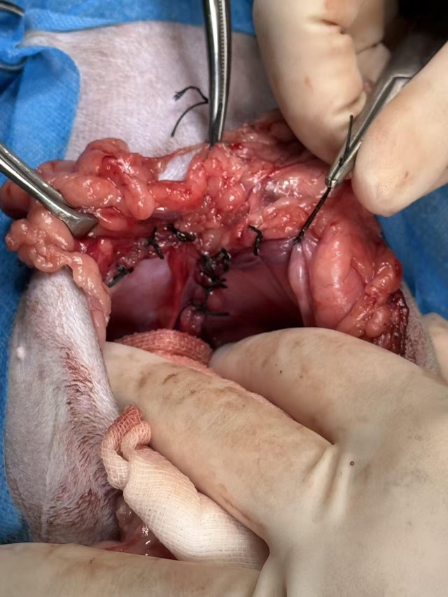
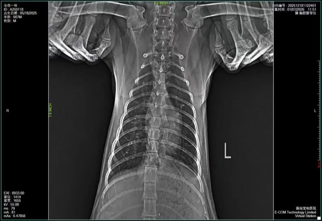
Diaphragmatic Hernia Repair
X-ray showed abundant abdominal-organ shadows in the thoracic cavity with compressed heart and lungs, confirming diaphragmatic hernia: large-area defect and rupture of the diaphragm. The defect was precisely measured and a custom-cut, slightly larger biosynthetic mesh fully covered and repaired it; X-ray at 12 days post-op showed good recovery and the patient was discharged.
Thoracic Cavity Repair
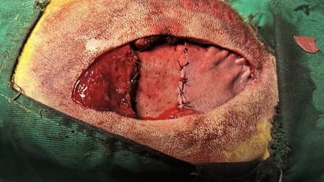
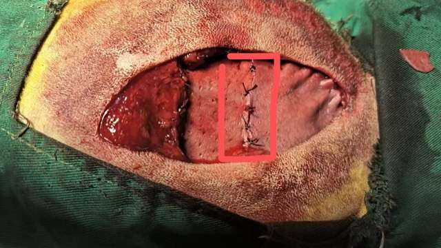
Bite wounds on both rib sides created two holes that soft tissue could not suture; hernia mesh was used for repair. As the area was too large, two pieces were pieced together; where the seam leaked, another mesh piece was overlaid and sutured in a “patching” fashion, with good post-op recovery.
Artificial Skin · Corneal Patch · Dura Mater Patch · Neurosurgery
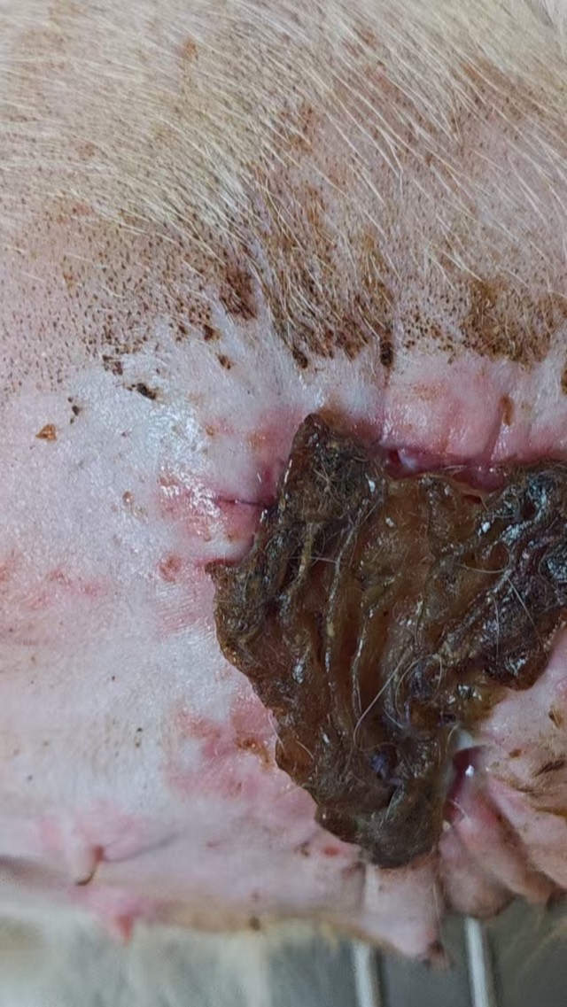
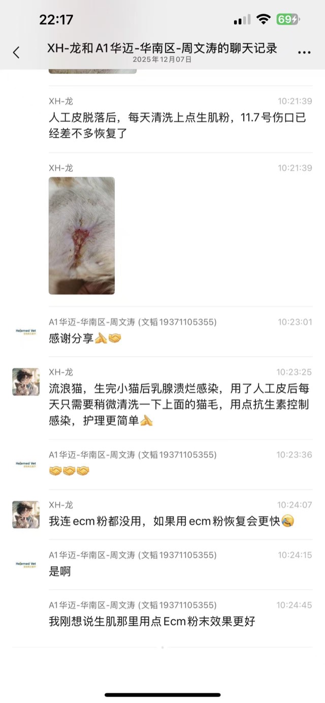
Artificial Skin for Skin-Defect Repair
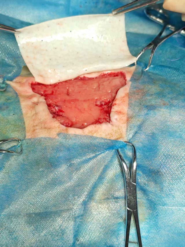
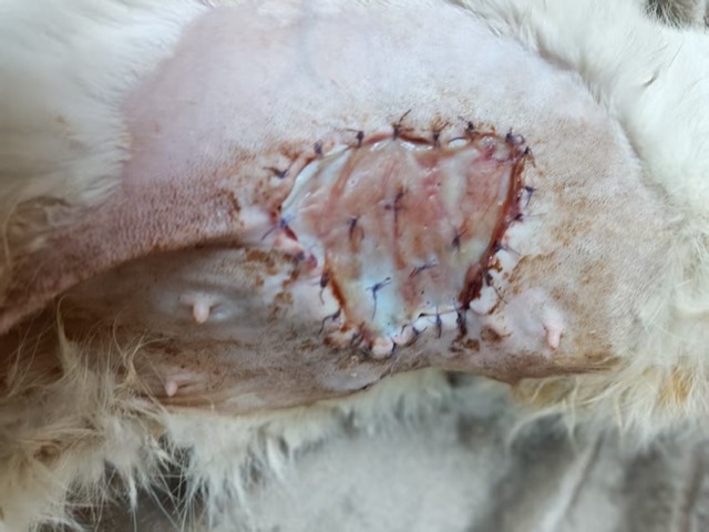
A stray cat developed mammary ulceration and infection after giving birth; after Biosynthetic Artificial Skin grafting, only light daily cleaning and antibiotics were needed — simple care. The artificial skin sloughed off naturally on Oct 30 and the wound basically recovered by Nov 7.
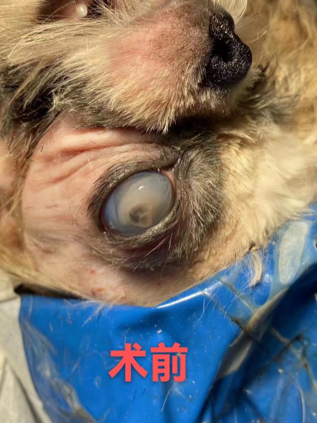
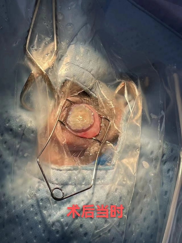
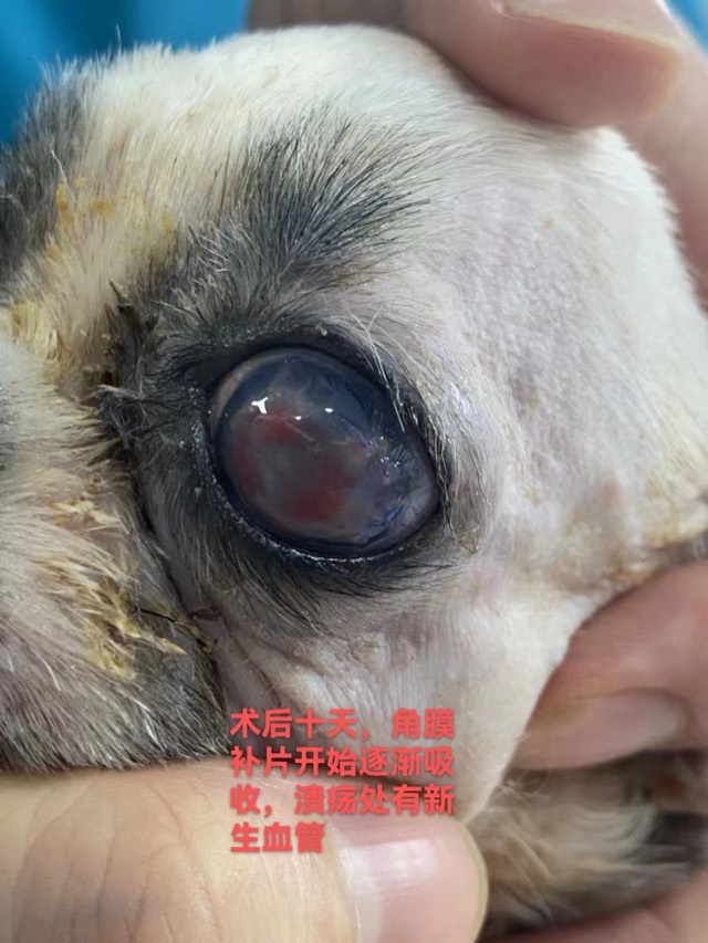
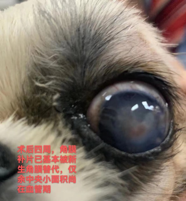
Corneal Patch Transplantation
The ECM membrane sheet (Corneal Patch) is used for corneal-layer repair such as ulcers and perforations: as an inductive scaffold for the corneal layer, recruiting cells and vessels to grow in and inducing corneal regeneration.
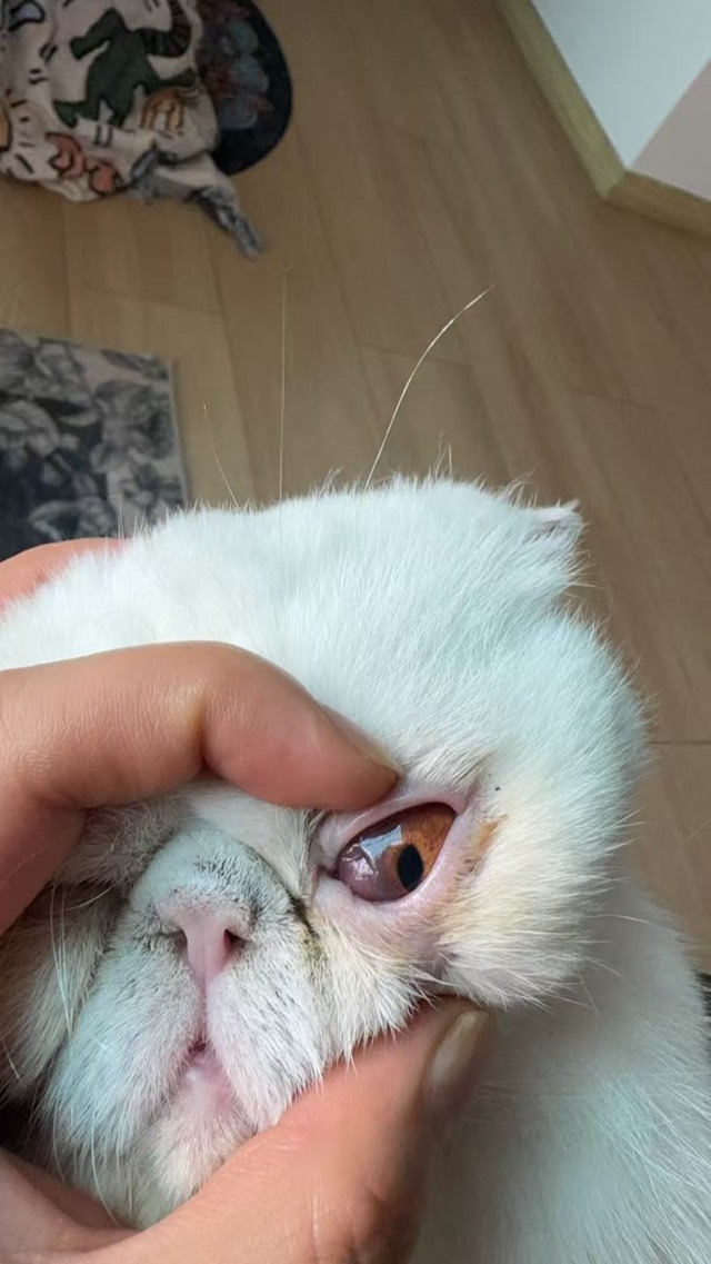
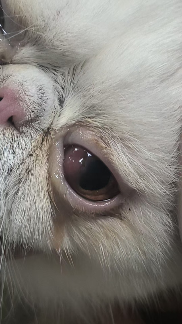
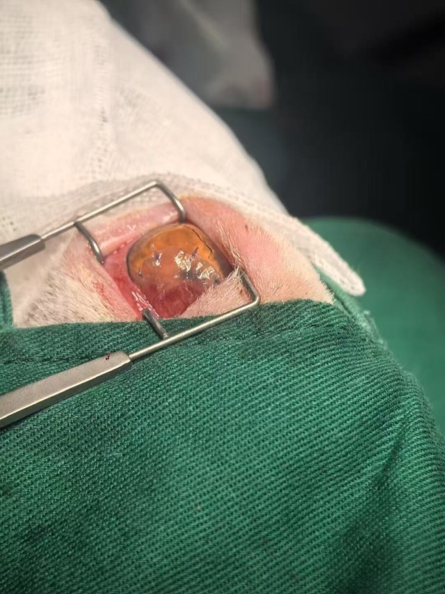
Corneal Patch Transplantation
Corneal repair surgery on Apr 27, 2025: the Corneal Patch's rough side faced the eyeball and smooth side outward; feedback on May 15 said corneal growth was very good, better than conjunctival-flap coverage.
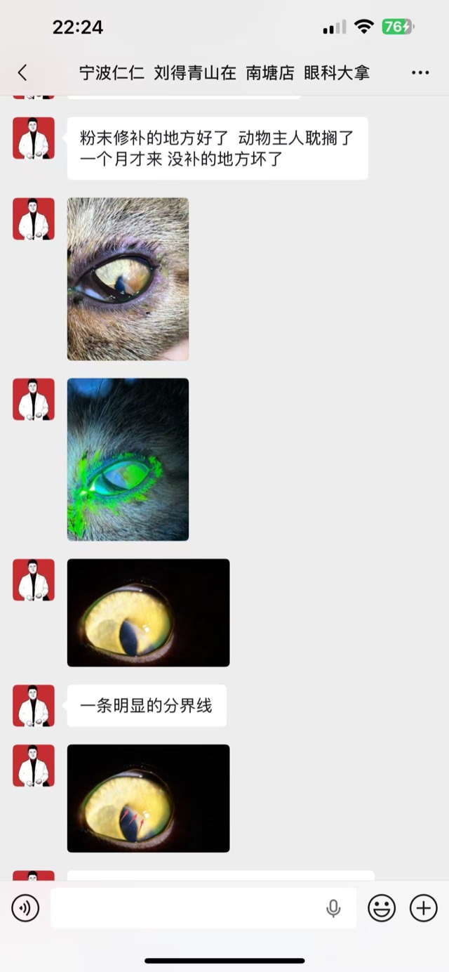
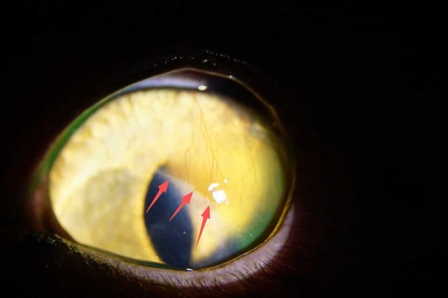
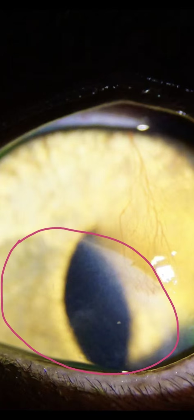
ECM Antibacterial Powder Corneal Application
ECM Antibacterial Powder was applied on the corneal layer with a bandage contact lens and eyelid suturing for protection; the covered area fully healed with a clear boundary line from the untreated area (outside the red circle is the untreated area).
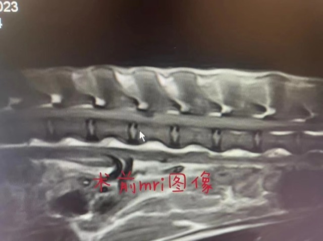
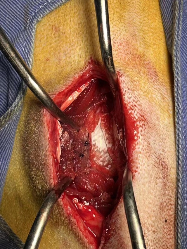
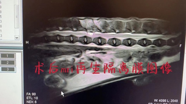
Hemilaminectomy (Disc Fenestration)
During neurosurgical hemilaminectomy, the ECM membrane sheet (Regeneration Barrier Membrane) was applied as a barrier and regeneration scaffold to protect neural tissue.
* The case images and treatment descriptions above are compiled from clinical sharing by partner hospitals. Individual differences are significant; please follow your surgeon's guidance for specific application plans.
Why Choose ECM Regenerative Materials?
- ECM Bioscaffold Technology — Based on extracellular matrix, retaining natural bioactivity and guiding tissue regeneration rather than permanent replacement.
- Full-Range Antibacterial Design — ECM Antibacterial Powder and the Antibacterial Artificial Bone series combine infection inhibition and regeneration induction.
- Complete Product Range — From ECM Cavity-Filling Sponge and ECM Tumor Filling Material to limb-bone bioregeneration material, alveolar bone filling material, Biosynthetic Absorbable Hernia Mesh and Corneal Patch — all in one place.
- For Hospitals — Prices are hospital prices, with online inquiry order submission supported.
Submit Inquiry Order
Fill in the information below and submit; we will confirm your order and shipping as soon as possible. You can also email directly to ecmvet@gmail.com or call +86 189 1832 1818。
⚡ Reply within 1 business day · 🔒 Info used for business contact only · 🚚 Worldwide shipping Submit Inquiry Order Now →