
- 1. Why an eye problem cannot wait
- 2. What "eye infection" turns out to be
- 3. Signs, and the ones that mean go now
- 4. What never to put in a dog’s eye
- 5. How a vet examines an eye
- 6. How each cause is treated
- 7. When infection damages the cornea
- 8. Corneal repair and the materials used
- 9. Clinical cases
- Frequently asked questions
- Related guides
1. Why an eye problem cannot wait
Three reasons, and they apply to every dog with a red or painful eye:
- The cornea has no blood supply of its own. It is nourished by tears and by fluid from inside the eye. That means it cannot mount the rapid inflammatory response that skin or muscle can, and infection or injury can progress to a deep ulcer in a day or two.
- A deep ulcer can perforate. Once the cornea is perforated, the fluid inside the eye leaks out, the eye collapses, and the options narrow sharply — often to surgical repair, and sometimes to removal of the eye. A surface ulcer treated on day one is a minor problem; a perforated eye is a major one.
- Pain in dogs is under-read. A dog will not complain. Squinting, rubbing the eye along the carpet, holding the eye closed, and reluctance to be touched near the head are all strong pain signals, and they appear late relative to the damage.
Go today if you see any of these: the eye is held closed · the cornea looks cloudy, blue or white · the dog is pawing at the face · the eye is bulging · the pupil looks different from the other eye · there is blood in the eye · the dog is suddenly bumping into things or reluctant to go out at night. These are not "wait and see" signs.
2. What "eye infection" usually turns out to be
Owners say "infection"; vets tend to say the specific diagnosis, because the treatment is different for each and several of these are not infections at all:
| Diagnosis | What it looks like | Key point |
|---|---|---|
| Conjunctivitis | Red, thickened eyelid lining, watery or mucoid discharge, mild squinting. The eye itself is clear. | The most common cause of a red eye. Usually bacterial or allergic, sometimes viral — the treatment differs and drops alone often fail if an underlying cause is present. |
| Keratoconjunctivitis sicca (dry eye) | Thick, sticky, yellow discharge, a dull cornea, recurrent "infections", often both eyes. | Not an infection — too few tears. Treated with tear stimulants and lubricants. Missed dry eye is why some dogs have "an eye infection" every few months. |
| Corneal ulcer | Squinting, watering, a rough or cloudy patch on the cornea, aversion to light. | An injury, not an infection, though infection follows. Confirmed with a fluorescein stain. |
| Blepharitis | Crusted, swollen eyelid margins, sometimes with hair loss around the eye. | Often part of a generalised skin problem, including allergy or a mite infestation. |
| Uveitis | A cloudy eye, a small pupil, a red eye with a deep ache, sensitivity to light. | Inflammation inside the eye. It needs systemic treatment, not eye drops alone, and it is often a sign of disease elsewhere in the body. |
| Foreign body or entropion | Constant tearing and rubbing, a rolling-in eyelid, a grass seed hidden under the third eyelid. | No amount of drops will help an eye that has a foreign body or eyelashes rubbing on it. |
| Cherry eye (prolapsed gland) | A red, fleshy mass in the inner corner of the eye. | Common in young dogs of certain breeds. Not an infection; needs surgical replacement of the gland. |
The overlap is what makes self-diagnosis unreliable. A dog with dry eye gets recurrent conjunctivitis; a dog with conjunctivitis rubs the eye and creates an ulcer; an ulcer becomes infected and turns from a grey patch into a melting cornea. Each step has a different treatment, and giving the wrong one first — particularly a steroid drop for what turns out to be an ulcer — is how the damage accelerates.
3. Signs, and the ones that mean go now
Signs you act on the same day:
- Cloudiness or a blue-white haze over the cornea — this is oedema or cellular infiltration, and it is a deep problem, not a surface one
- Yellow-green or thick purulent discharge, rather than the clear watery discharge of a simple irritation
- The eye is kept shut, or the dog squints constantly
- Pawing at the face, or rubbing the eye on furniture and carpet
- A visible defect on the surface of the eye — a rough, dull or pitted patch, or a dark spot
- Swelling or bulging of the eyeball, or an eye that looks different in size from the other
- Blood inside the eye, a torn or irregular pupil, or a sudden loss of vision
Signs worth an appointment this week rather than today:
- Intermittent watering, or crusting in the inner corner each morning, with a dog that otherwise seems comfortable
- A red eye with no discharge and no squinting — often allergy, or an early dry eye
- Recurrent "eye infections" that come back every few months, which usually means the real problem is dry eye, a lid deformity, or an allergy
4. What never to put in a dog’s eye
This section matters more than any other in this guide. The eye is small, poorly supplied with blood, and unforgiving:
- Never use a steroid eye drop (or a combination antibiotic-and-steroid drop) before a fluorescein stain has been done. Steroids suppress the repair response in the cornea. On an ulcer they cause melting — the cornea dissolves — and the most common route to a perforated eye in dogs is a steroid drop given for an ulcer that nobody stained for.
- Do not use leftover human eye drops from your own medicine cabinet. Human preparations have different concentrations, different preservatives, and some are simply not for use in animals. Drops prescribed for your own eye, or for another dog, are not a solution to this eye.
- Do not use a home-made saline or salt solution as treatment. Flushing an eye with a sterile eye wash to remove debris is reasonable; making your own solution and instilling it repeatedly is not, because it is neither sterile nor isotonic.
- Do not use human antibiotic ointment intended for skin on the eye — the base is not designed for the cornea and can cause a chemical injury.
- Do not try to remove a foreign body that is embedded in the cornea, or push a protruding mass back in. You can flush gently with sterile saline while waiting, and that is all.
- Do not use a cone as a substitute for treatment. An Elizabethan collar is a very useful part of treatment — it stops self-trauma — but it does not treat the infection, and it does not protect a perforated eye.
If you have already applied a drop you now suspect was wrong, do not wait to see what happens. Take the bottle with you and tell the vet exactly what was put in, how much, and when. That single piece of information changes how the eye is managed.
5. How a vet examines an eye
An eye examination is quick, painless, and almost all of it is done in the consulting room:
- Fluorescein stain — a yellow dye that sticks to exposed corneal tissue. This is how an ulcer is confirmed, and it is the test that must be done before any steroid is considered.
- Schirmer tear test — a paper strip in the lower lid measures tear production in 60 seconds. It is how dry eye is found.
- Tonometry — measures the pressure inside the eye, for glaucoma.
- Slit lamp or magnified examination — assesses the depth of an ulcer and checks for a foreign body under the eyelids.
- Cytology and culture — a swab or a scrape identifies bacteria or yeast and their sensitivity, which is what to culture before reaching for antibiotics.
- Blood tests — for uveitis, since the inflammation inside the eye is often driven by infection or immune disease elsewhere.
6. How each cause is treated
- Conjunctivitis. Antibacterial drops or ointment if bacterial, antihistamines or anti-inflammatories if allergic, and treatment of the underlying cause. A conjunctivitis that keeps coming back is a symptom, not a diagnosis; the vet looks for dry eye, a lid deformity, distichiasis (extra eyelashes) or allergy.
- Dry eye. Tear stimulants (ciclosporin or tacrolimus), lubricant drops and gels, and sometimes an antibiotic for the secondary infection at the start. This is a lifelong condition that needs continuous medication — stopping treatment because the eye looks better is followed by relapse.
- Corneal ulcer. An antibiotic drop to control infection, pain relief, and in many cases a contact lens or a temporary tarsorrhaphy (partially suturing the eyelids shut) to protect the surface while it heals. The dog wears a cone continuously. The ulcer is re-stained at intervals to confirm it is shrinking.
- Blepharitis. Treatment depends on the cause — antibacterial or antifungal medication, mite treatment, or management of the underlying allergy. Cleaning the lid margins is part of it.
- Uveitis. Anti-inflammatory treatment, often systemically, plus atropine to relieve the painful spasm of the pupil, and treatment of the underlying disease.
- Foreign body or entropion. The mechanical problem is removed — the grass seed out, the lid corrected surgically. Medical treatment alone will not fix an eye that is being physically rubbed.
- Cherry eye. Surgical replacement of the gland into its normal position. Removing the gland instead is no longer standard practice, because the gland produces a substantial part of the tear film and its loss causes dry eye.
Whatever the cause, two things are always part of the treatment: pain control, because pain drives rubbing and rubbing drives damage; and an Elizabethan collar, because the moment the dog is comfortable it will try to scratch the eye that is healing.
7. When infection damages the cornea
An ulcer that is not controlled progresses in a predictable way, and the stages matter because they change the surgery:
| Stage | What is happening | Usual management |
|---|---|---|
| Superficial ulcer | Loss of surface epithelium only; the cornea is still basically intact | Medical treatment, protection, recheck. Most heal in a week to ten days |
| Stromal (deep) ulcer | The ulcer has eaten into the body of the cornea; the eye may look cloudy and be very painful | Aggressive medical treatment, and surgery may be needed to support the surface |
| Melting ulcer | Collagenases from bacteria and from the cornea itself are actively digesting the cornea | A true emergency. Intensive treatment and surgery; the cornea can perforate within hours |
| Descemetocele | Only Descemet’s membrane is left, a thin transparent sheet; the eye bulges under the pressure | Surgery now — a graft, a flap or a membrane is used to hold the eye together |
| Perforation | The cornea has given way; fluid is leaking and the eye is collapsing | Emergency surgical repair, or enucleation if the eye cannot be saved |
This is the sequence in which a "minor eye infection" becomes an eye that has to be removed. Not every case follows it, and most do not — but the cases that do are the ones where treatment was delayed, or where a steroid was used on an unstained eye. That is why the advice in this guide is to have the eye looked at rather than treated blind.
8. Corneal repair and the materials used
When the cornea has lost tissue — after a deep or melting ulcer, a perforation, or a sequestrum — it has to be physically reconstructed. Repair is done under general anaesthesia by a veterinarian, and the surgical options include:
- A conjunctival flap — a piece of the dog’s own conjunctiva is rotated over the defect and sutured in place, bringing a blood supply to the area. Robust and well established, but it is opaque, so the eye is covered while it heals.
- A corneal graft — donor cornea, or a preserved biological membrane, used to replace or support the lost layer.
- A biological scaffold patch — a thin, absorbable sheet placed on the defect, which acts as a barrier and as a scaffold that the corneal layer grows into. Because it is thin and transparent in use, it is an option for repairs where the conjunctival flap would be too bulky, and it degrades as the cornea regenerates.
For the scaffold type, two practical points matter. The rough side faces the eyeball and the smooth side faces outward — the orientation is not arbitrary, and it is decided by the surgeon. And the patch protects the defect while the cornea heals underneath; it does not replace the need for the antibiotic and pain treatment that goes with it, or for the cone.
Materials used in corneal repair
Our ECM regenerative materials are veterinary surgical materials used by the treating veterinarian after the eye has been assessed and stained. The Bio-Guided Regeneration Membrane is used as a corneal patch — the 20×20mm single-layer is the thinnest in the series for milder defects, and the 20×30mm double layer, which degrades more slowly and has greater tensile strength, is used for more severe repair such as perforation, sequestrum, deep ulcer and established infection. ECM Antibacterial Powder has also been applied to the corneal layer under a bandage contact lens and with eyelid suturing for protection.
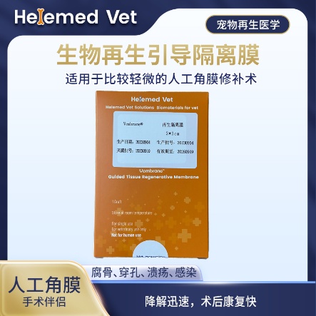
Bio-Guided Regeneration Membrane (2×2cm)
Corneal patch: for milder artificial corneal repair (sequestrum, perforation, ulcer, infection); fast degradation and quick post-op recovery, so it is also the thinnest regenerative isolation membrane in the series.
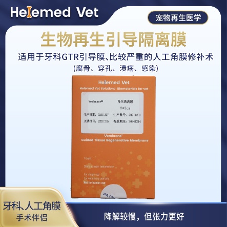
Bio-Guided Regeneration Membrane (2×3cm)
Double-layer GTR isolation membrane and GBR barrier membrane — a regenerative barrier membrane for dental guided tissue regeneration (GTR) and guided bone regeneration (GBR), plus more severe artificial corneal repair (sequestrum, perforation, ulcer, infection); slower degradation but better tension.
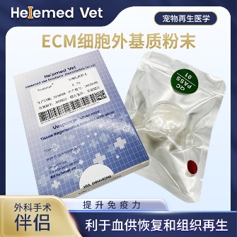
ECM Antibacterial Powder
ECM bioscaffold powder — an extracellular matrix (ECM) based antibacterial and inhibitory powder: widely used as ECM trauma dressings on infected internal/external wounds, fresh wounds, burns and crush injuries to markedly improve blood supply, accelerate healing, inhibit infection and reduce complications; it is also an ECM hemostatic material that stops capillary bleeding. How to use: after debridement, take a small amount of powder with forceps and apply to the wound surface, moisten with saline (moist wound healing) and bandage; apply once every 5 days — daily debridement is not required.
Supplied to veterinary clinics and hospitals. These are surgical materials used by the treating veterinarian — they are not eye drops and cannot be used at home.
9. Clinical cases
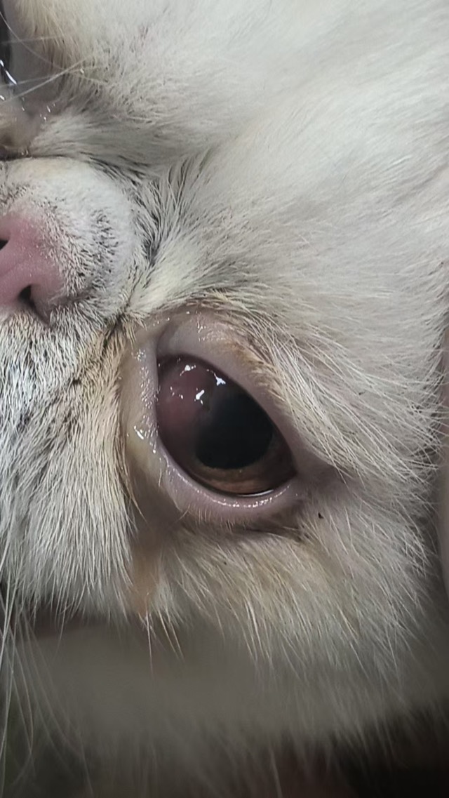
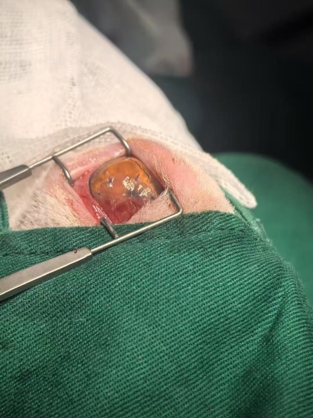
Three cases from our partner hospitals, all of them eyes that had already lost tissue: a corneal patch transplant where the corneal layer regenerated, a surgical repair using the patch, and an antibacterial powder application to the cornea protected by a contact lens and eyelid sutures.
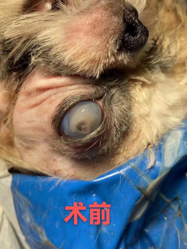
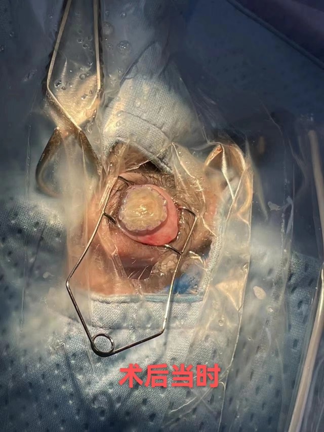
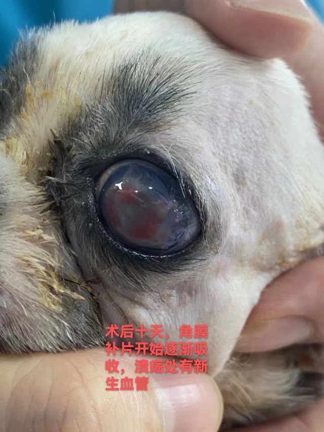
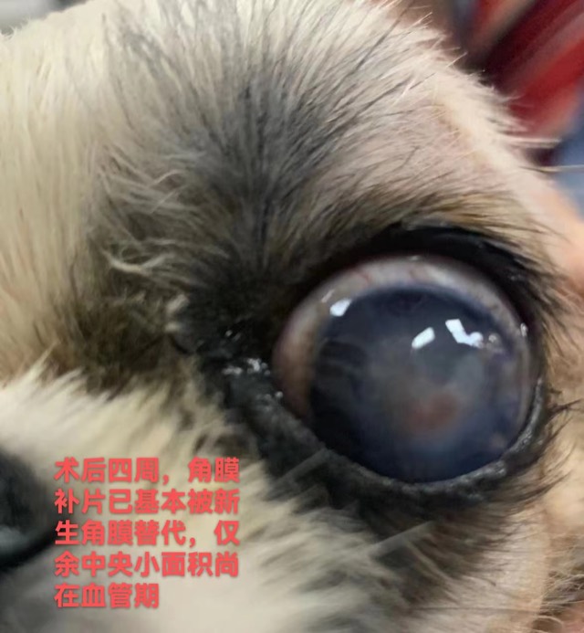
Corneal Patch Transplantation — Corneal Recovery
The ECM membrane sheet (Corneal Patch) is used for corneal-layer repair such as ulcers and perforations: as an inductive scaffold for the corneal layer, recruiting cells and vessels to grow in and inducing corneal regeneration.
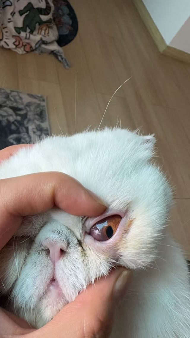
Corneal Patch Transplantation — Surgical Repair
Corneal repair surgery on Apr 27, 2025: the Corneal Patch's rough side faced the eyeball and smooth side outward; feedback on May 15 said corneal growth was very good, better than conjunctival-flap coverage.
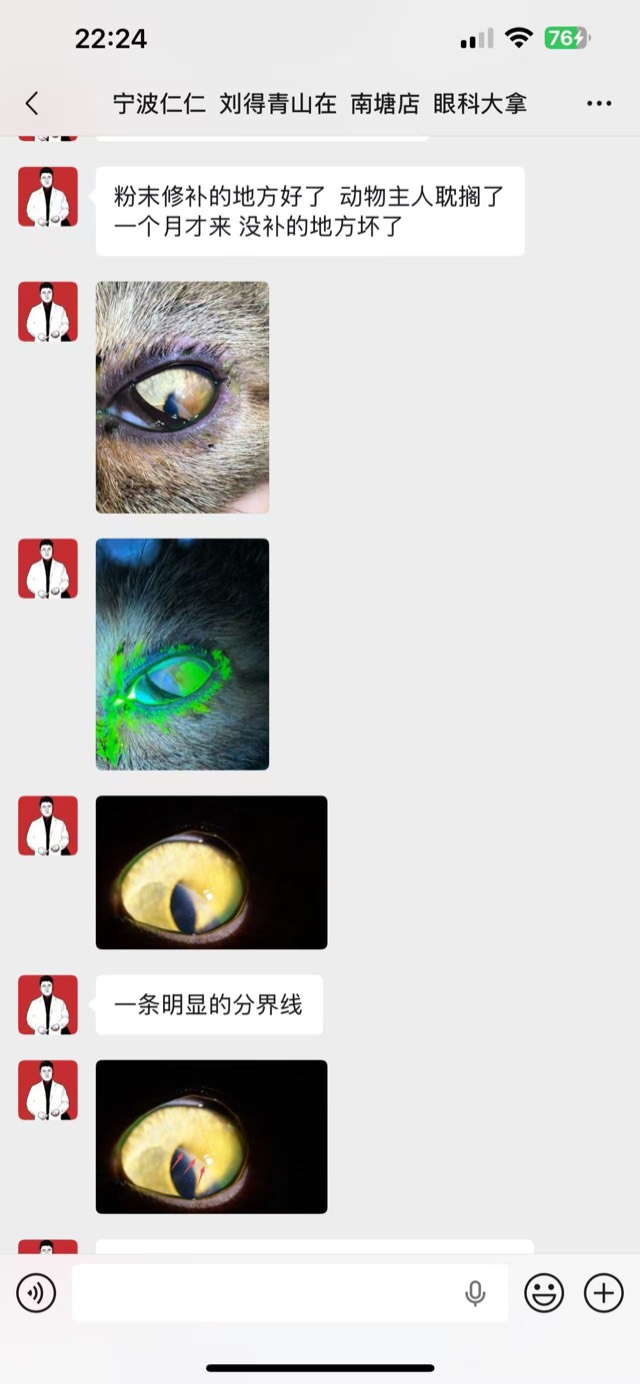
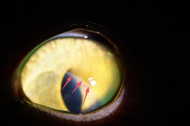
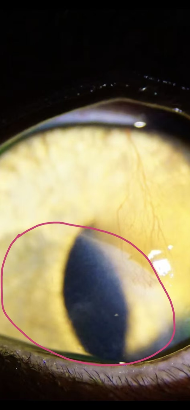
ECM Antibacterial Powder Corneal Application
ECM Antibacterial Powder was applied on the corneal layer with a bandage contact lens and eyelid suturing for protection; the covered area fully healed with a clear boundary line from the untreated area (outside the red circle is the untreated area).
Cases from partner veterinary hospitals. ECM materials are veterinary surgical materials and are applied by the treating veterinarian. See all 39 clinical cases →
Frequently asked questions
How can I tell if my dog’s eye infection is serious?
Signs that mean the dog should be seen today: the eye is kept closed or constantly squinting, the cornea looks cloudy, blue or white, the dog paws at the face, the eye bulges, the pupils look different from each other, or there is a visible defect on the eye surface. Watering with a clear discharge and a comfortable dog is usually less urgent, but still needs an appointment.
Can I use human eye drops on my dog?
No — and one group is genuinely dangerous. A steroid or antibiotic-and-steroid drop given on an ulcer can cause the cornea to melt and perforate. Never use drops prescribed for yourself or for another dog, and never use a drop containing a steroid before a vet has done a fluorescein stain to rule out an ulcer.
What causes a dog eye infection?
Bacteria and, less often, viruses or yeast, but usually there is an underlying reason: dry eye, an allergy, a foreign body such as a grass seed, an eyelid that rolls in, extra eyelashes, or a corneal injury. That is why an eye infection that keeps coming back usually means the underlying cause was never treated.
How is a dog eye infection treated?
It depends on the diagnosis. Bacterial conjunctivitis is treated with an antibacterial drop; dry eye with tear stimulants and lubricants, lifelong; an ulcer with antibiotic drops, pain relief, protection and a cone; uveitis with systemic anti-inflammatory treatment. In every case the cone matters, because rubbing re-injures the eye.
Why does my dog keep getting eye infections?
Recurrent eye problems in dogs are most often dry eye (keratoconjunctivitis sicca), an eyelid or eyelash abnormality, or an allergy. A Schirmer tear test takes a minute and finds the most common of these. Repeated courses of drops without a diagnosis is the usual reason the problem keeps coming back.
What happens if a corneal ulcer is left untreated?
It deepens. A superficial ulcer that heals with treatment can progress to a stromal ulcer, then a melting ulcer where the cornea is actively digested, then a descemetocele where only a thin membrane is left, and finally a perforation where the eye collapses. Perforated eyes often cannot be saved, so the cost of waiting is high.
Can a damaged cornea be repaired?
Often yes, if the eye is treated in time. A veterinarian may use a conjunctival flap, a graft, or a thin absorbable scaffold patch that supports the surface while the corneal layer regenerates under it. The outcome depends on how much tissue has been lost and whether the inside of the eye is still intact.