
- 1. Why most dog teeth are extracted
- 2. Which teeth, and what changes when they go
- 3. What happens during the extraction
- 4. The socket problem
- 5. What goes wrong if it is left alone
- 6. Bone grafting and guided regeneration
- 7. Oronasal fistula: the complication to prevent
- 8. Recovery at home, day by day
- 9. Clinical cases
- Frequently asked questions
- Related guides
1. Why most dog teeth are extracted
The overwhelming majority of tooth extractions in dogs are the end point of periodontal disease, and the mechanism is worth understanding because it explains why the surrounding bone is involved at all:
- Plaque forms on the tooth surface within hours of cleaning.
- Plaque mineralises into tartar, which is visible as the brown deposit on the tooth. By itself, tartar does not cause the damage — it is a shelf that collects more plaque.
- The gums become inflamed (gingivitis). At this stage the process is reversible with a professional clean.
- The attachment is destroyed (periodontitis). The supporting structures — gum, periodontal ligament and alveolar bone — are progressively lost. This stage is not reversible, and pockets form between tooth and gum where bacteria multiply.
- The tooth becomes mobile, painful or infected, or the bone loss leaves the root exposed. At this point, saving the tooth may require advanced periodontal surgery, and extraction is often the right decision — particularly in a dog with several affected teeth.
Other reasons for extraction: a fractured tooth with exposed pulp, a tooth that has died and gone dark, severe wear, a tooth involved in a tumour resection, retained deciduous (baby) teeth that are crowding the adult teeth, and teeth damaged in a trauma. Dental disease severe enough to need extraction is also not only a mouth problem — chronic periodontal infection is a constant bacterial load on the kidneys, liver and heart.
2. Which teeth, and what changes when they go
| Tooth | Why it is extracted | What matters afterwards |
|---|---|---|
| Incisors (front, small) | Periodontal disease; they work loose early because they have single roots and thin bone | Minor functional loss — mostly used for grooming and nibbling. Alveolar bone loss here affects the shape of the front of the jaw. |
| Canines (the large fangs) | Fracture, periodontal disease with deep pockets, trauma | The most significant extraction in the dog. They anchor the tongue, guide the bite, and their roots are long and curved — the upper canine root sits close to the nasal cavity, which is why this is the extraction most likely to create an oronasal fistula. |
| Premolars | Periodontal disease; the upper fourth premolar is commonly affected | Used for shearing and for holding food. Loss is usually tolerated well. |
| Carnassials (upper 4th premolar and lower 1st molar) | Severe periodontal disease, fracture, or a tooth that has split | The main shearing teeth. Extracting them changes how the dog chews; the opposing tooth will be trimmed or removed to prevent it overgrowing. |
| Molars | Severe periodontal disease; molars have multiple roots and often need sectioning | The upper molars sit just under the eye, and an infected molar is a classic reason for a swollen face or a swelling below the eye. |
Two consequences owners rarely expect. First, removing one tooth often changes the opposing one: with nothing to wear against, the opposing tooth continues to erupt and can grow into the gap, so the vet may plan to extract or trim it at the same time. Second, the jaw behind an extracted tooth loses bone, and this is progressive and irreversible unless the socket is managed properly at the time of extraction.
3. What happens during the extraction
A modern extraction is a surgical procedure, not a tug:
- General anaesthesia with local nerve blocks. Dental work in dogs is done under full general anaesthesia, with local anaesthetic blocks to control pain. Cleaning or extracting teeth in a conscious dog is neither safe nor humane, and it is not how it should be offered to you.
- Full-mouth dental radiographs. Two thirds of the tooth is below the gum. X-rays show the roots, the bone level, and pathology that cannot be seen from the surface — including teeth that look fine and are not.
- Closed extraction for a tooth with a single, straight root and enough bone: the tooth is loosened and elevated out.
- Surgical (open) extraction for multi-rooted teeth, curved roots and the canines: a flap of gum is raised, bone may be removed with a bur (alveolectomy) to expose the roots, and the tooth is sectioned into single-rooted pieces before removal. This is the standard for the teeth most likely to be done badly.
- Socket debridement. The socket is cleaned out, the thin bone of the socket wall is smoothed, and any remaining diseased tissue is removed.
- Suturing and, where indicated, bone grafting. The gum is closed and, for the sockets that need it, a graft material is placed before closure.
- Post-operative radiograph. A picture after extraction confirms no root fragment was left behind — the most common cause of a socket that does not heal.
Retained root fragments deserve a specific mention: root tips left in the jaw keep the inflammation going, they can migrate, and they cause a chronically draining tract that owners notice as a bad smell or a recurring swelling months later. The post-operative radiograph is what prevents this.
4. The socket problem
An extraction leaves a hole in the bone called an alveolar socket. What happens to that hole over the following weeks is the part of dentistry owners are least often told about:
- The socket fills with a blood clot, which is the natural scaffolding for healing.
- Granulation tissue and new bone form, if the socket is protected and there is no infection.
- But the bone remodels inward. Without support, the wall of the socket collapses as it heals, and the ridge narrows — the bone that supported the tooth is not replaced at its original dimensions.
In humans the consequence is a ridge that is too thin for an implant later. In dogs the consequences are different and more immediate: a crater that traps food, an uneven ridge where the gum sits lower than on the neighbouring teeth, less bone around the remaining teeth, and — for the upper teeth whose roots sit against the nose — a socket wall thin enough to break through into the nasal cavity.
This is the reason modern veterinary dentistry treats the socket, not just the tooth: the point of a graft in an extraction site is to preserve the bone around it, so the ridge stays solid, the gum stays in place over it, and the neighbouring teeth keep their support.
5. What goes wrong if it is left alone
- Oronasal fistula — a permanent connection between the mouth and the nasal cavity, most often after upper canine extraction. Food and liquid come out of the nose, and the dog sneezes and has nasal discharge when it eats. It does not close by itself.
- Bone loss and a sunken ridge, which weakens the jaw and removes the support from the neighbouring teeth
- Jaw fracture — a real risk in small breeds where the mandible is thin and most of the bone around the lower canine is tooth root. Extraction in these dogs, and the management of the socket, needs care.
- Root fragment retained, causing a chronic draining tract, bad breath and pain
- Delayed or failed healing of the gum, with the socket left open and exposed bone
- Food impaction in the defect, which is uncomfortable and keeps the area inflamed
6. Bone grafting and guided regeneration
When a tooth is extracted, the options are to close the gum over the empty socket and accept the bone loss, or to reconstruct the site. Reconstruction goes by two related names:
- Alveolar bone reconstruction / site preservation — placing a bone graft or scaffold material into the socket to support the ridge and induce new bone formation, then closing the gum over it.
- Guided tissue regeneration (GTR) and guided bone regeneration (GBR) — using a barrier membrane over the defect to keep fast-growing soft tissue out while slower-growing bone and periodontal tissue regenerate beneath it. GTR is used to attempt to save a tooth with a periodontal defect; GBR is used to rebuild bone, for example around an extraction site or a defect.
The steps are standard and the sequence is what makes them work:
- Clean and debride. No graft tolerates infected or necrotic tissue in the bed. The socket is curetted and irrigated until the walls are clean and bleeding.
- Fill the defect with the graft or scaffold material, packing it into the socket so it is in contact with the bone walls.
- Cover it — a barrier membrane over the graft where GTR or GBR is being used, held in place so soft tissue cannot invade the space.
- Close the soft tissue over the site with a tension-free suture line. This is the step that most often decides success: a closure under tension opens, and an open closure means the graft is lost and the defect is exposed again.
Which material is used depends on the defect. Granule graft materials are used to fill alveolar sockets and periodontal defects after extraction; larger granule sizes are more economical for multiple extractions. A double-layer membrane, which degrades slowly and has more tensile strength, is used as the barrier over GTR and GBR sites.
Why this is worth doing at the time, and not later: once the ridge has collapsed and the gum has healed over it, reconstructing the site means re-opening the gum, re-creating the defect and grafting into a scarred bed. Doing it at the time of extraction is simpler, cheaper and more predictable.
7. Oronasal fistula: the complication to prevent
An oronasal fistula is a hole between the oral and nasal cavities. It is the complication that veterinary dentists warn about most, because it is common, it is avoidable, and it does not heal on its own.
Two situations cause it. The first is advanced periodontal disease around an upper canine, where the bone between the tooth root and the nasal cavity has already been destroyed — the hole was there before the extraction, and the extraction reveals it. The second is extraction technique: if the socket is not closed properly over a thin or damaged wall, or a flap is closed under tension, the suture line gives way and leaves a permanent defect.
Signs: food or water coming out of the nose while eating, sneezing fits during meals, nasal discharge, a bad smell from the nose, and a nose that is irritated on one side only. Owners often describe it as "the dog sneezes milk after drinking".
Treatment is surgical. The defect is reopened, the tissue is freshened, and the site is closed in layers using a graft to fill the defect and a flap to cover it — the same logic as any other bone defect. Repair is more likely to succeed at the first attempt than after repeated failures, which is why the priority is prevention at the time of extraction.
8. Recovery at home, day by day
| Days | What to expect | What you do |
|---|---|---|
| Day 0–1 | The dog is drowsy from anaesthesia, may have a little blood-tinged saliva, and may whine or be unsettled. Swelling of the muzzle is common after multiple extractions. | Offer water in small amounts, a light meal in the evening if the vet allows, keep the dog warm, quiet and indoors. No stairs or jumping. |
| Days 2–4 | Mouth discomfort peaks, then improves. Some dogs are reluctant to eat hard food. | Soft food: tinned food, soaked kibble, or a veterinary recovery diet. Give pain relief exactly as prescribed — do not adjust or replace it yourself. |
| Days 5–10 | Gums look pink and clean. Sutures loosen and are usually dissolvable. | Continue soft food, no chewing on toys, no bones, no sticks. Keep the cone on if one was sent home. Check the mouth daily for a bad smell or a swelling. |
| Weeks 2–4 | The socket is filling in and the gum has healed. | Return for the recheck. Ask the vet to look at the extraction site rather than deciding for yourself that all is well. |
Call the clinic rather than waiting if you see: bleeding that does not stop, a swelling that grows, a bad smell from the mouth, discharge from the nose or food coming out of the nose when eating, the dog refusing all food for more than 24 hours, or drooling far more than expected. A swelling under the eye after a dental procedure needs to be seen the same day.
Do not give human painkillers. Paracetamol and ibuprofen are toxic to dogs. If the prescribed pain relief seems inadequate, call the vet that same day.
One last point that is easy to forget: extraction relieves pain, but it does not cure the disease. Periodontal disease has no cure — it has management. The remaining teeth still need home brushing, and professional cleaning under anaesthesia at the interval the vet recommends, or the same process starts again in the teeth that are left.
Materials used in alveolar bone reconstruction
Our ECM regenerative materials are veterinary surgical materials used by the treating veterinarian. The antibacterial artificial bone is an alveolar bone filling material used for periodontal regeneration and alveolar bone reconstruction after extraction — it fills the socket, induces new bone formation, works as a hemostatic material and provides a sustained antibacterial effect. The double-layer regeneration membrane is the barrier used over dental GTR and GBR sites.
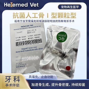
Antibacterial Artificial Bone Type I (Granules)
Alveolar bone filling material for dental applications, periodontal regeneration, alveolar bone reconstruction after extraction and bone-graft (ECM bone transplanted materials) procedures; induces autologous bone regeneration, works as a hemostatic material, inhibits infection and fills defects for a bone-graft effect.
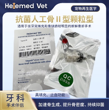
Antibacterial Artificial Bone Type II
Large-size dental ECM artificial bone material and alveolar bone filling material: periodontal regeneration, GBR guided bone regeneration and alveolar bone reconstruction after extraction; more economical for multiple teeth and repeated surgeries.
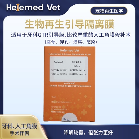
Bio-Guided Regeneration Membrane (2×3cm)
Double-layer GTR isolation membrane and GBR barrier membrane — a regenerative barrier membrane for dental guided tissue regeneration (GTR) and guided bone regeneration (GBR), plus more severe artificial corneal repair (sequestrum, perforation, ulcer, infection); slower degradation but better tension.
Supplied to veterinary clinics and hospitals. Ask your veterinarian whether site preservation is appropriate for your dog’s extraction.
9. Clinical cases
Three cases from our partner hospitals: tooth extraction with alveolar bone reconstruction and the standard four steps of rebuilding the ridge, an oronasal fistula filled and closed, and guided tissue regeneration used to save a tooth with a periodontal defect rather than extracting it.
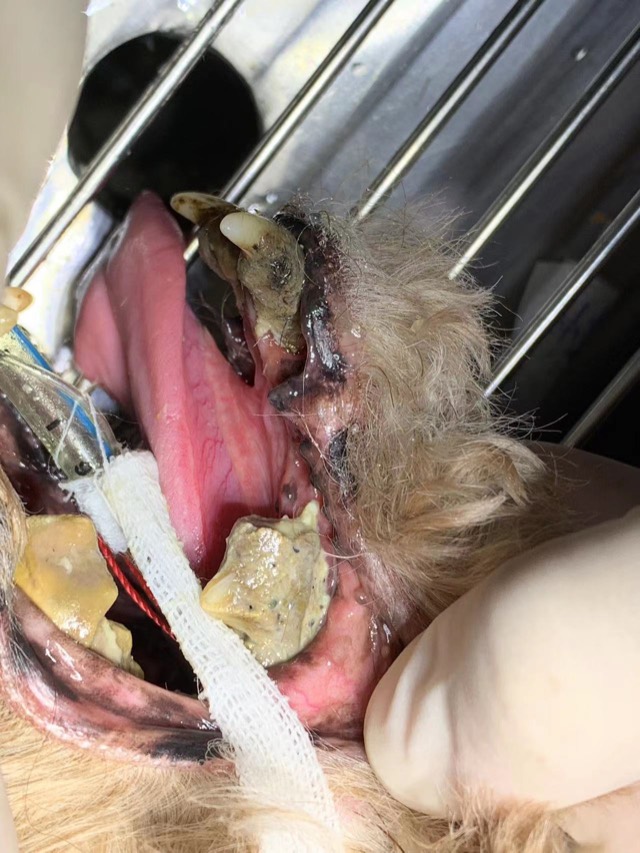
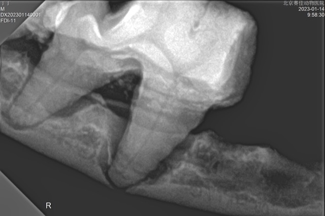
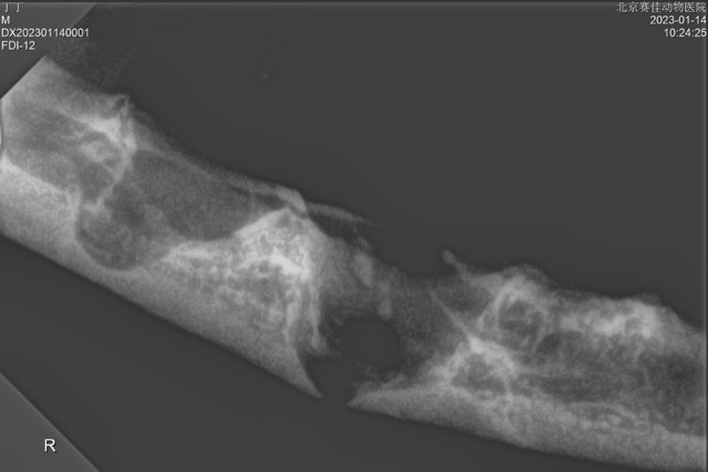
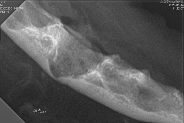
Tooth Extraction and Alveolar Bone Reconstruction
After extraction, site preservation surgery is needed: alveolar bone regeneration and alveolar bone reconstruction prevent fracture, oronasal fistula, facial fistula and other risks. X-ray showed mandibular dissolution and defect; after filling with Antibacterial Artificial Bone granules (alveolar bone filling material) the alveolar bone was regenerated. No infection or fracture post-op, and re-check showed good mandibular regeneration.

Oronasal Fistula Filling and Hemostasis
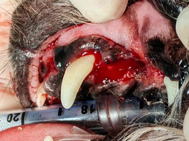
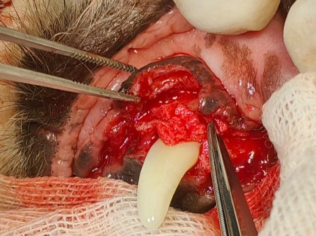
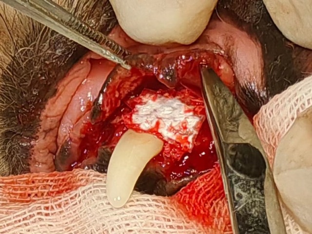
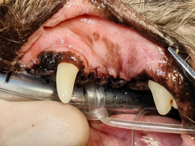
Oronasal fistula occurs not only in unextracted cases but also commonly after extraction — improper suturing leaves a hidden risk, and the same defect can open into a facial fistula. If the cavity is large and bleeding is heavy during mucosal flap reflection, cut ECM Sponge to size and pack the fistula tract, then do a flap suture, hooking the sponge with a few stitches to prevent inhalation into the nasal cavity. The sponge fully grows into soft tissue and blocks the fistula.
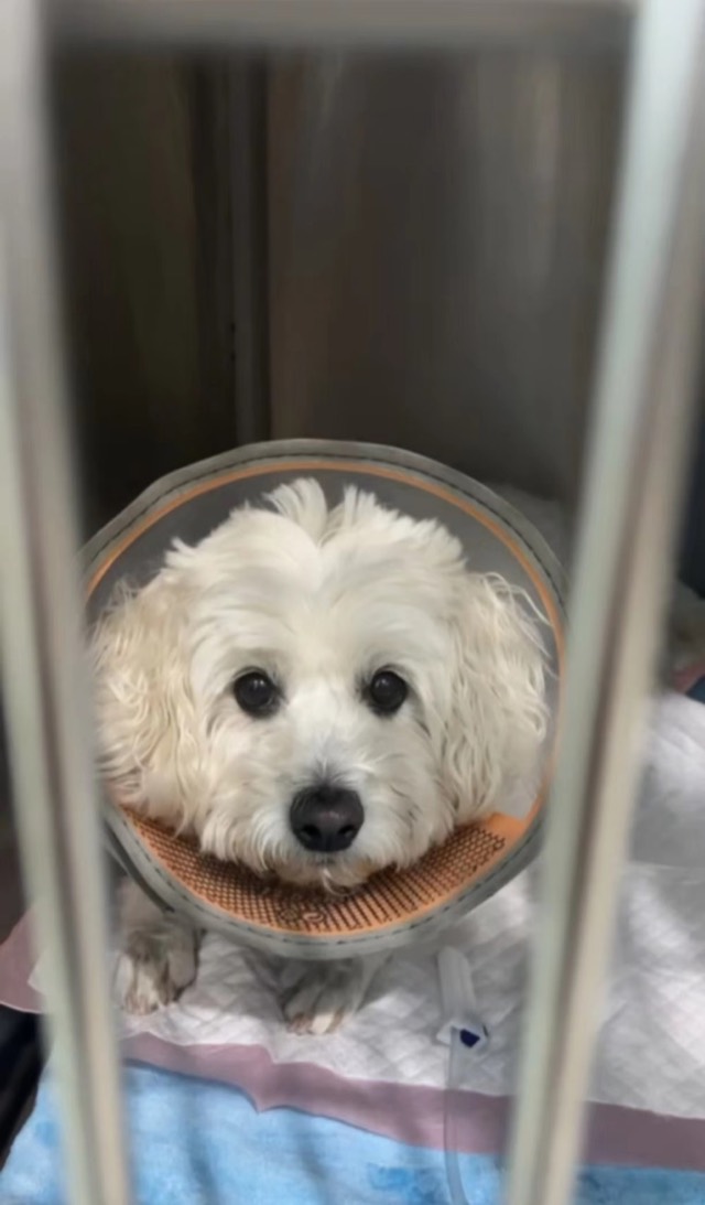
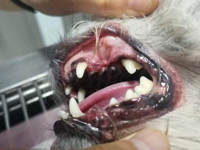
Periodontal GTR Guided Regeneration (Tooth-Saving)
A 10-year-old dog with severe periodontal disease — swollen and inflamed gums and alveolar bone atrophy; the owner declined multiple extractions and chose GTR tooth preservation (GTR tooth-saving surgery): mucosal flap reflection, extraction of the periapical-periodontitis tooth, root planing and repeated curettage, Antibacterial Artificial Bone granules packed and covered with Regeneration Barrier Membrane (smooth side inward), tension-free nodular suturing, and “button” principle to tighten the periodontal space around the canine. The goal of the procedure was periodontal regeneration, subgingival regeneration and periodontal tissue regeneration at the same site. At 10-day suture removal tooth stability was better, with good follow-up.
Cases from partner veterinary hospitals. ECM materials are veterinary surgical materials and are applied by the treating veterinarian. See all 39 clinical cases →
Frequently asked questions
How long does it take a dog to recover from a tooth extraction?
The mouth is usually comfortable enough for normal soft eating within three to five days, and the gum closes over the socket in one to two weeks. The bone underneath fills in over several weeks. The anaesthetic often takes more out of an older dog than the extraction does, so a quiet day or two afterwards is normal.
How many teeth can be removed at once?
As many as are diseased. It is common for a dog with advanced periodontal disease to have ten or more teeth removed in a single anaesthetic, including both sides of the mouth. Doing it in one procedure means one anaesthetic and one recovery, and it is kinder than a series of visits.
Will my dog still be able to eat normally after extractions?
Yes. Dogs use their teeth less for chewing than people assume — they tear and swallow. Most dogs with few or no teeth manage dry kibble perfectly well, and many eat more comfortably afterwards than before, because the diseased teeth were painful. Soft food is used during the healing period, then the diet is usually returned to normal.
What is an oronasal fistula and how do I know if my dog has one?
It is a hole between the mouth and the nose, usually after an upper canine extraction. The signs are food or liquid coming out of the nose when the dog eats, sneezing during meals, nasal discharge and a bad smell on one side. It does not close by itself and needs surgical repair.
Do I need to do anything about the empty socket?
That is the conversation to have with your vet before the extraction. Left alone, the socket heals but the surrounding bone collapses inward, which weakens the ridge and the support of the remaining teeth — and in the upper jaw a thin socket wall can open into the nose. Grafting the socket is done to prevent that, and it is easiest at the time of extraction.
Should my dog have dental X-rays before an extraction?
Yes. Most of the tooth and all of the bone are under the gum. Radiographs show the root shape, the bone level, retained roots, and pathology that cannot be seen from the surface. A post-extraction radiograph also confirms that no root fragment has been left behind — the most common reason a socket fails to heal.
Is anaesthesia safe for an old dog having dental work?
Age itself is not the deciding factor — general health is. A pre-anaesthetic blood panel, sometimes chest X-rays and an ECG, and a tailored anaesthetic protocol make dental work feasible for most older dogs. Leaving painful infected teeth untreated carries its own risk, including kidney, liver and heart strain from chronic infection.