
- 1. What an ear hematoma is
- 2. Why it happens
- 3. What it looks like
- 4. Will it go away on its own?
- 5. Treatment options, compared honestly
- 6. What the operation involves
- 7. Aftercare: drains, bandages and collars
- 8. Why the ear can crumple afterwards
- 9. Preventing the next one
- 10. Clinical cases
- Frequently asked questions
- Related guides
1. What an ear hematoma is
The ear flap — the pinna — is not a single piece of tissue. It is two sheets of cartilage with a thin layer of tissue, blood vessels and connective fibres sandwiched between them. Those two sheets are normally held together tightly enough that there is no space between them at all.
An aural hematoma is what happens when a blood vessel inside that sandwich breaks. Blood is pumped into the space between the two cartilage sheets, and because the sheets can separate, it accumulates there. The result is a pocket of blood, a fluid-filled swelling that can cover part of the ear or the whole flap.
The word itself just means a collection of blood outside a blood vessel. The important detail is where it collects: in a space that is not supposed to exist, and that the body cannot easily drain or fill in.
2. Why it happens
Mechanically, this is not a mystery. A dog shakes its head hard — and dogs can shake their heads with real force — and the acceleration shears the small vessels between the cartilage sheets until one tears. Some dogs will do it repeatedly for hours.
What makes a dog shake its head that hard is the more useful question, because if it is not identified, the hematoma comes back:
| Cause | Why it leads to a hematoma |
|---|---|
| Ear infection (otitis externa) | The most common single cause. An infected ear is itchy and painful, and the dog shakes and scratches at it relentlessly. |
| Environmental or food allergy | Ears are skin. Allergic dogs commonly have itchy ears, and recurrent ear disease is one of the classic signs of an allergy that has not been identified. |
| Ear mites | Intensely itchy; common in puppies and in households with cats. |
| Foreign body | A grass awn or seed in the canal drives sudden, violent head shaking, often after a walk. |
| Polyps or tumours in the canal | Irritation and discharge keep the ear uncomfortable. |
| Skin disease of the ear flap | Any itchy or inflamed condition of the flap itself can start the cycle. |
| Trauma | A bite, a knock, or rough handling of an already painful ear. |
There is also a recognised association with certain blood vessel and clotting disorders, and with some medications, but these are uncommon. In the overwhelming majority of dogs, the answer is an uncomfortable ear.
3. What it looks like
- A swelling of the ear flap, from a small blister to the entire ear. It looks like the ear has been inflated.
- Soft and squishy to the touch — described as fluctuant. Unlike an abscess, which is usually hot and painful, a fresh hematoma is often surprisingly painless and feels like a water-filled cushion.
- It appears fast — typically within hours of the dog starting to shake its head.
- The ear may droop or sit at an odd angle, because of the weight and the swelling.
- The dog may shake, tilt or scratch — a continuation of the behaviour that caused it.
- The ear canal may be dirty, red or smelly, which is your clue to the underlying cause.
An abscess of the ear flap feels different: firmer, hotter, and generally painful. A tumour feels solid and stays put rather than appearing overnight. If the swelling is not soft and it did not appear suddenly, the diagnosis is probably not a simple hematoma.
4. Will it go away on its own?
Technically, yes, and this is where owners are often given incomplete advice. If nothing is done:
- The blood slowly reabsorbs over two to four weeks.
- The two cartilage sheets heal back together, but they heal with fibrous scar tissue between them, because the space was never closed and the surfaces were never kept in contact.
- The result is a thickened, crumpled, permanently deformed ear flap — sometimes called a cauliflower ear. It is cosmetic rather than dangerous, but it is irreversible.
- During those weeks the ear is uncomfortable, the dog may keep shaking it, and the pocket can re-bleed, become infected, or organise into a hard lump.
So the honest position is: leaving it alone is a legitimate choice for a small hematoma in an older dog where the appearance of the ear does not matter, provided the underlying ear disease is treated. For a large hematoma, a young dog, or an owner who cares about the ear's shape, it is not the best option.
5. Treatment options, compared honestly
| Option | What it involves | Trade-off |
|---|---|---|
| Do nothing | Treat the underlying ear disease and let the blood resorb | Slow; almost always leaves a thickened, crumpled ear; can re-bleed or become infected |
| Needle drainage alone | The fluid is drawn out with a needle and syringe | Relief lasts days at best. The space is still there, so it simply refills. High recurrence rate and not a treatment |
| Drainage plus a steroid injection | The fluid is drained and a long-acting corticosteroid is injected into the space to suppress inflammation and the re-accumulation of fluid | Can work in small, fresh hematomas, sometimes with repeated injections. Less reliable in large ones, and the ear may still deform |
| Surgery — incision, clot removal and sutures | The pocket is opened, the clot and fibrous material are removed, and the two cartilage sheets are stitched together with a pattern of sutures so the surfaces heal as one layer | The most reliable way to restore the ear's shape, and the standard treatment for a large hematoma. Requires anaesthesia and a period of aftercare, and the ear may still not be perfectly flat |
| Surgery with a drain | As above, with a drain left in place for a few days so fluid cannot accumulate | Very effective; needs drain care and a bandage, and the drain is removed at a recheck |
| Drainage holes (window technique) | A series of small openings are made in the flap so the pocket cannot hold fluid | Effective in some cases and leaves less suture line, but healing takes longer and the ear requires careful dressing |
Whatever route is chosen, one step is not optional: the ear canal has to be examined and the cause treated. A perfectly repaired hematoma in a dog with an untreated ear infection will very often be followed by another one.
6. What the operation involves
- Anaesthesia and preparation. The ear is clipped and surgically prepared. The dog is anaesthetised — this is genuinely painful surgery and it cannot be done properly on a struggling patient.
- Opening the pocket. An incision is made along the inner surface of the flap, over the length of the swelling, and the accumulated blood is released.
- Removing the clot and fibrous material. The old blood has often organised into a clot and a layer of fibrin. This is removed, and any dead tissue with it, so that two clean surfaces are left facing each other.
- Closing the dead space. This is the part that decides the outcome. The two cartilage sheets are sutured together across the whole area of the pocket, so that no space is left for fluid to collect in, and the surfaces are held in contact while they heal. Small openings may be left or a drain placed so that any further fluid has somewhere to go.
- Dressing and support. A bandage is applied to hold the ear in the right position and to protect the suture line. The ear may be supported against the head rather than left hanging.
- Treating the ear canal. While the dog is anaesthetised, the canal is examined properly and cleaned, and appropriate treatment started. This is the step that prevents the next hematoma.
Two things decide how the ear ends up looking: how completely the dead space was closed, and how the ear is managed in the days afterwards.
7. Aftercare: drains, bandages and collars
- The collar goes on and stays on. A dog that can reach the ear can undo the surgery within minutes. A collar, or for some dogs a head bandage, is worn continuously until the vet says otherwise.
- Keep the bandage dry and check it daily. If it slips, gets wet, or smells, call the vet — a wet bandage over a fresh surgical site is a recipe for infection.
- Expect some swelling. A degree of post-operative swelling and bruising is normal. Rapidly increasing swelling, a fresh collection of fluid, or a discharge needs a recheck.
- The drain, if placed, is removed at a recheck — usually after a few days. Do not remove it yourself.
- Keep treating the ear canal as prescribed, and finish the course.
- Ask about pain relief and give it as directed. A comfortable dog shakes its head less.
- Return for the recheck even if everything looks fine. Suture lines and ear canals are both easy to misjudge at home.
Where the surgery has left a cavity or a deeper defect — which happens when a hematoma has organised, or when a mass or a diseased canal has been removed at the same time — the wound also has to be managed as a wound: infection controlled, dead tissue removed, dead space dealt with, and the surface covered while it heals.
Materials used where a surgical cavity has to be filled
Our ECM regenerative materials are veterinary surgical materials, applied by the treating veterinarian at the time of surgery. ECM Sponge is used to fill and support a surgical cavity — the space left once a hematoma, a mass or a diseased ear canal has been removed — while the antibacterial powder is applied to the debrided wound bed.
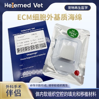
ECM Sponge
ECM filling sponge and ECM cavity-filling biomaterials: for internal soft-tissue cavity filling, providing long-lasting space occupation and inducing regeneration to achieve a soft-tissue graft effect; also an ideal ECM Tumor Filling Material for dead-space repair after tumor resection, and a routine companion to ECM trauma dressings in cavity and fistula wounds.
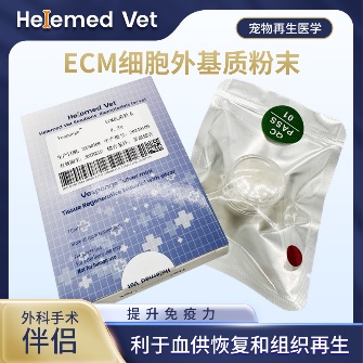
ECM Antibacterial Powder
ECM bioscaffold powder — an extracellular matrix (ECM) based antibacterial and inhibitory powder: widely used as ECM trauma dressings on infected internal/external wounds, fresh wounds, burns and crush injuries to markedly improve blood supply, accelerate healing, inhibit infection and reduce complications; it is also an ECM hemostatic material that stops capillary bleeding. How to use: after debridement, take a small amount of powder with forceps and apply to the wound surface, moisten with saline (moist wound healing) and bandage; apply once every 5 days — daily debridement is not required.
Ask your veterinarian whether these materials suit your dog's surgical site; they are supplied to veterinary clinics and hospitals.
8. Why the ear can crumple afterwards
A thickened, folded or crumpled ear after a hematoma is common, and it is worth understanding because it is not bad luck. It happens when:
- The space was never closed, so the body filled it with fibrous scar tissue between the two cartilage sheets.
- Sutures came out or were removed too early, letting the surfaces separate again.
- The dog shook or scratched the ear during healing and disrupted the repair.
- The hematoma was large or long-standing, so there was more organised clot and more fibrosis.
- The hematoma recurred, adding a second round of scar tissue.
A small amount of thickening is normal and usually acceptable. Marked crumpling can be prevented far more easily than it can be corrected afterwards, which is the strongest practical argument for treating the underlying ear disease early and following the aftercare instructions precisely.
9. Preventing the next one
- Treat the ear disease properly, including the full course of treatment and the recheck. This is the single most important measure.
- Find out why the ear was itchy. In adult dogs, recurrent ear disease is usually a symptom of an allergy, and an allergy that is never worked up produces ear after ear after ear.
- Check the ears regularly — weekly, and after every swim or bath. Look at the canal, not just the flap, and notice any smell.
- Dry the ears after water contact, particularly in breeds with heavy, pendulous ears.
- Keep flea control up all year, on every pet in the house.
- Act on a swollen ear immediately. A hematoma seen the day it appears is easier to manage than one that has been organising for a week.
10. Clinical cases
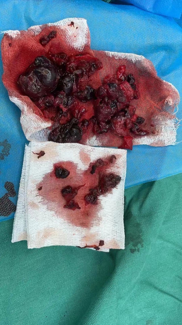
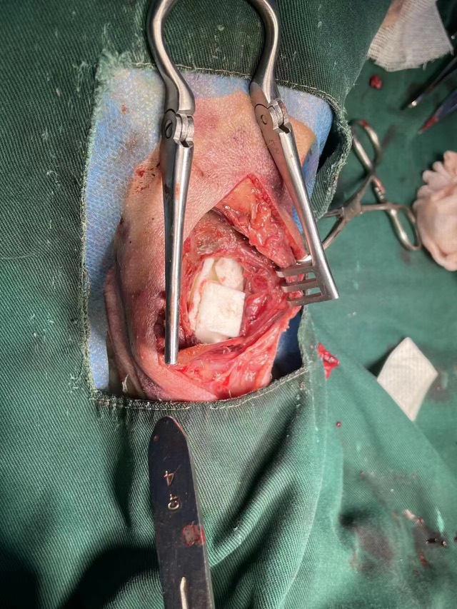
These two cases from our partner hospitals show ECM Sponge used to fill a surgical cavity left after tissue has been removed — the same problem a hematoma operation creates, where the space left behind has to be supported and closed while the ear heals.
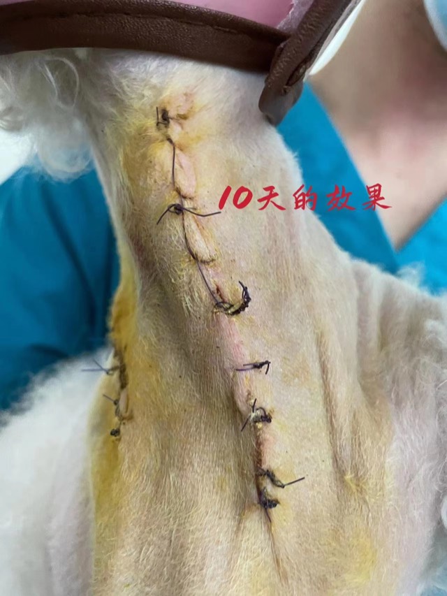
ECM Sponge Assisted Cavity Filling
When there is an obvious cavity with normal surrounding blood supply, ECM Sponge can assist filling (larger volume, ~20-day degradation, synchronized with new tissue). Before use, fully soak in saline and squeeze out excess water.
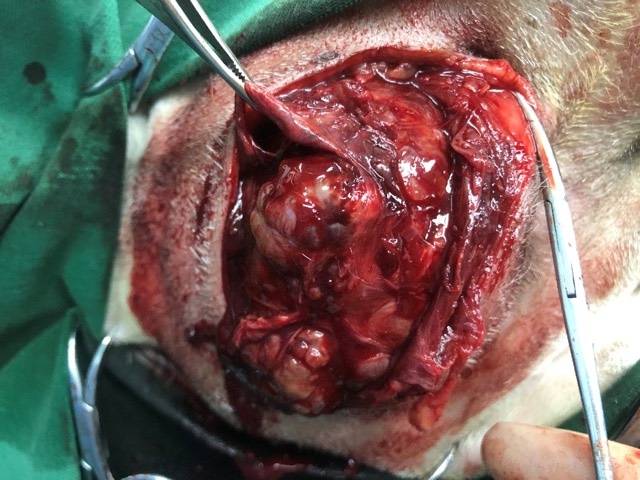
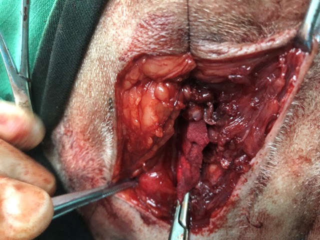
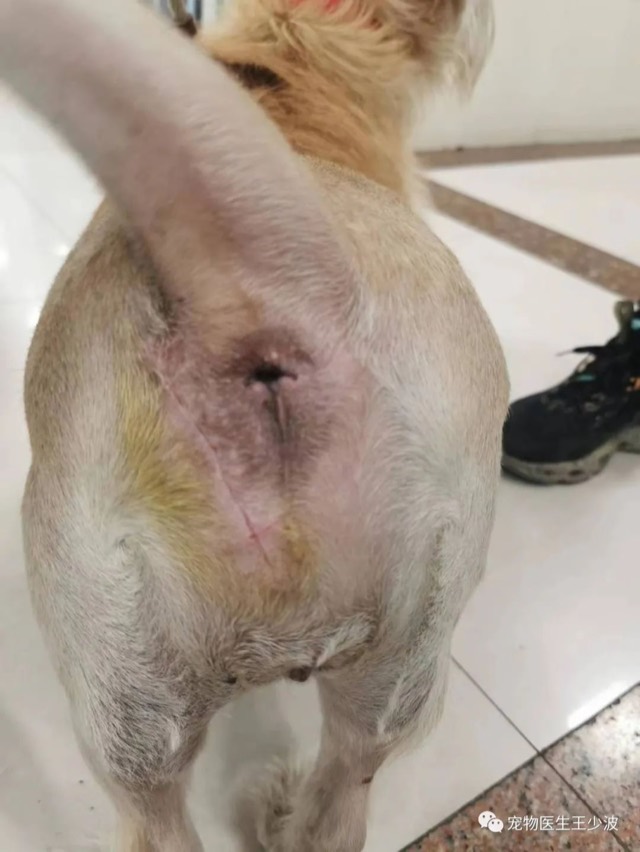
Tumor-Resection Cavity Filling
Tumor-resection surgery often has poor post-op healing; where an obvious cavity exists, implant ECM Sponge for filling (like tissue grafting), lay ECM powder subcutaneously for prophylaxis, then close the wound — effectively accelerating primary healing. Two pieces of ECM Sponge were implanted in this case.
Cases from partner veterinary hospitals. ECM materials are veterinary surgical materials and are applied by the treating veterinarian, not at home. See all 39 clinical cases →
Frequently asked questions
What does an ear hematoma look like on a dog?
A soft, swollen, balloon-like ear flap that squishes when touched. It usually appears within hours of the dog shaking its head, and it can affect part of the flap or all of it. Unlike an abscess it is often not particularly hot or painful.
Will a dog ear hematoma go away on its own?
The blood is gradually reabsorbed over two to four weeks, but the ear usually heals with scar tissue between the cartilage layers, leaving a thickened, crumpled ear. It is a reasonable choice for a small hematoma in some dogs, but the underlying ear disease still has to be treated.
Is ear hematoma surgery necessary?
Not always, but it is the most reliable way to restore the ear shape, and it is the standard treatment for a large hematoma. Small, fresh hematomas are sometimes managed by drainage with a steroid injection. Draining with a needle alone is not a treatment, because the space refills.
What causes an ear hematoma in dogs?
Almost always violent head shaking or scratching, caused by an uncomfortable ear — most often an ear infection, an allergy, ear mites, or a foreign body in the canal. The hematoma is the symptom; the ear problem is the cause.
How long does it take to recover from ear hematoma surgery?
The sutures typically stay in for around two to three weeks, and the bandage period is usually shorter than that. The ear looks swollen for several days and then settles. Full healing of the cartilage takes longer. Follow the recheck schedule rather than judging it yourself.
Can I drain my dog’s ear hematoma at home?
No. Draining it with a needle removes fluid, not the pocket, and it will refill — usually within days — while introducing infection into a closed space. Any procedure on the ear needs to be done by a vet, under appropriate sedation or anaesthesia.
Does a dog ear hematoma need to be seen urgently?
It is not usually a life-threatening emergency, but it should be seen promptly — within a day or two. The sooner it is assessed, the more options are available and the less organised the clot becomes, which makes the surgery smaller and the outcome better.