
- 1. Why most dogs have it by age three
- 2. The four stages, and the one that reverses
- 3. Signs owners miss
- 4. Why the mouth is not only a mouth problem
- 5. What a professional dental cleaning involves
- 6. What actually works at home
- 7. Saving the tooth, or extracting it
- 8. Clinical cases
- Frequently asked questions
- Related guides
1. Why most dogs have it by age three
Periodontal disease is inflammation and destruction of the tissues that hold a tooth in its socket: the gum, the periodontal ligament and the alveolar bone. It begins the same way in every dog:
- Plaque forms on the tooth surface within hours of a meal. It is a soft, sticky biofilm of bacteria, and it is invisible while it is thin.
- Plaque mineralises into tartar — the brown or yellow deposit along the gum line. Tartar is not itself destructive; it is a rough shelf that collects more plaque and holds it against the gum.
- The gum becomes inflamed. This is gingivitis: red, swollen, bleeding gum margins. At this stage the damage is entirely reversible with a professional clean and effective home care.
- The attachment is destroyed. Once the inflammation moves below the gum line into the periodontal ligament and bone, the disease is periodontitis. Attachment and bone that are lost do not grow back on their own, and the process continues under a gum line that often still looks pink.
Three facts explain why it is so common. Dogs do not brush their teeth, so plaque is never removed between cleanings. Dogs are long-lived now, so years of accumulation are the norm rather than the exception. And small and flat-faced breeds are crowded: teeth are packed together with abnormal gaps where plaque collects, and the roots of the upper teeth sit close to the nasal cavity and under the eye, so disease there has consequences beyond the mouth.
The consequence owners are least prepared for is that the most damaged teeth often look the most normal from above the gum. Two thirds of the tooth, and all of the supporting bone, are below the surface. That is the part that has to be examined with radiographs rather than looked at.
2. The four stages, and the one that reverses
Veterinary dentistry grades periodontal disease by how much attachment has been lost, measured with a probe around each tooth and confirmed on radiographs:
| Stage | What is happening | What it needs |
|---|---|---|
| 0 — Healthy | No plaque or minimal plaque, pink gum margin, no attachment loss | Home care, and professional monitoring at the routine examination. |
| 1 — Gingivitis | Plaque and tartar at the gum line, gum margin red and swollen, may bleed on probing. No attachment loss. | Fully reversible. A professional clean under anaesthesia, then daily home care to stop it coming back. |
| 2 — Early periodontitis | Up to about 25% of attachment lost; early bone loss and shallow pockets visible on radiographs. | Professional clean, subgingival scaling, and home care. Usually still manageable, and the teeth can generally be kept. |
| 3 — Moderate periodontitis | 25–50% attachment loss, deeper pockets, gum recession, possible bleeding and discomfort when eating. | Periodontal treatment, which may include advanced procedures to save specific teeth. Some teeth will be better out than kept. |
| 4 — Advanced periodontitis | More than 50% attachment loss, bone loss deep enough to reach the end of the root, mobile teeth, pus in the pockets, sometimes a fistula into the nose. | Extraction of the affected teeth in most cases, plus treatment of the surrounding sites and the options in the sections below. |
Stage 1 is the only stage at which the disease is cured rather than controlled, and it is the stage most often missed — because a dog with gingivitis only has red gums and bad breath, and nobody looks. Everything after it is management: slowing the loss, keeping the remaining teeth supported, and preventing the complications.
3. Signs owners miss
Signs that are easy to dismiss and should not be:
- Bad breath. The commonest first sign, and the one most often put down to the dog being a dog. Smell near the gum line rather than in the middle of the mouth.
- Yellow-brown tartar along the gum line, particularly on the upper fourth premolar and the canines, where plaque collects first.
- Red, swollen or bleeding gums. Roll the lip up gently on both sides and compare the two sides of the mouth.
- Gum recession — roots that look exposed, teeth that appear to be getting longer.
- Chewing on one side, dropping food, or refusing hard food and chewing only on soft things. These are pain signs, and they are reliable.
- Reluctance to have the mouth touched, or pulling away when the muzzle is handled.
- Excessive drooling, or drooling with blood in it.
- Pawing at the mouth, or rubbing the muzzle along the ground.
Two signs that point specifically to advanced disease in the upper jaw, because the roots of the upper canines and premolars sit against the nasal cavity and under the eye:
- Nasal discharge or sneezing when eating, or food and water coming out of the nose — a sign that a tract has opened between mouth and nose (an oronasal fistula).
- A swelling below one eye, or a draining tract on the cheek — an infected tooth root presenting as a facial problem. This is a common reason owners take a dog to the vet for an eye or skin complaint and leave with a dental diagnosis.
Dogs hide dental pain well. A dog that is eating normally may still have several teeth that are painful when pressed, and the absence of a change in appetite is not evidence that the mouth is fine. If you can smell the mouth, or see tartar at the gum line, the disease is already past stage 0.
4. Why the mouth is not only a mouth problem
A periodontal pocket is not a sealed space — it is an open wound at the gum margin, colonised by hundreds of bacterial species, sitting directly on a rich blood supply. The consequences reach well beyond the mouth:
- Continuous bacteria in the bloodstream. Chewing, and even gentle probing, sends bacteria from the pocket into the circulation. In a healthy dog the body clears this; in a dog with a damaged heart valve or reduced kidney reserve, that chronic exposure matters.
- Kidneys and liver. Chronic periodontal infection is a persistent antigenic and inflammatory load on both, and renal changes are consistently found in dogs with advanced disease.
- Heart. Bacterial endocarditis and valvular disease are associated with chronic oral infection, particularly in small breeds already prone to valve disease.
- Diabetes. The relationship runs in both directions: diabetes worsens periodontal disease, and chronic oral inflammation makes blood glucose harder to control.
- Local destruction. The same process that loses bone allows a fistula into the nose, and in the lower jaw of a small breed it can weaken the mandible to the point of fracture.
This is the reason veterinary dentists push back on "just take out the bad teeth when they bother him". Managing the disease early is not cosmetic work; leaving it is a decision about the whole dog, made over years, usually without the owner realising a decision was being made.
5. What a professional dental cleaning involves
A proper dental procedure under anaesthesia is a series of steps, and most of them cannot be done in a conscious dog:
- A pre-anaesthetic assessment. A blood panel, and in older or unwell dogs imaging and an ECG, to choose an anaesthetic protocol safely. Age on its own is not a reason to refuse the procedure.
- Full-mouth dental radiographs. Two thirds of every tooth is below the gum. Radiographs show root shape, bone level, retained roots, resorptive lesions and pockets that are invisible from the surface — including teeth that look intact and are not.
- Probing and charting. Pocket depth is measured around every tooth and recorded, which is how the stage is assigned and how progress is tracked at the next visit.
- Subgingival and supragingival scaling. An ultrasonic scaler removes tartar above and, critically, below the gum line, where the disease actually is.
- Polishing. Scaling leaves microscopic scratches on the enamel; polishing smooths them so that new plaque does not cling to a roughened surface.
- Irrigation, and where indicated fluoride or a sealant.
- Extraction, periodontal treatment, or referral, depending on what the chart and the radiographs show. This is decided during the procedure as often as before it.
- A recovery plan with a written home-care protocol, because the procedure buys a clean mouth and home care decides how long it stays clean.
On "anaesthesia-free dental cleaning": scaling a conscious dog removes only what can be seen above the gum line, which is the least important part, and leaves the subgingival plaque and tartar that drive the disease entirely in place. The mouth looks better and the disease continues — and with an uncooperative patient there is a real risk of injury to the gum and to the scaler. It is not a substitute, and it is not a cheaper version of the same thing.
6. What actually works at home
Home care decides how often the dog needs to go back under anaesthesia, so it is worth being specific about what works and what does not:
| Approach | Verdict | Notes |
|---|---|---|
| Daily tooth brushing | The single most effective measure | A soft pet toothbrush with pet toothpaste. Daily is the target; several times a week still helps. Never use human toothpaste — fluoride and xylitol are toxic to dogs, and the foaming agent is not meant to be swallowed. |
| Veterinary dental chews and diets | Useful as an addition | Products with a texture that mechanically cleans the tooth surface and carries a recognised veterinary oral health claim are worth choosing. They support brushing; they do not replace it. |
| Antiseptic rinses, gels and water additives | Helpful where brushing is impossible | A reasonable second line for dogs that will not tolerate a brush, and useful in the weeks after a dental procedure. |
| Bones and hard chews | Do not use | Bones and chews hard enough to resist a fingernail break teeth, and fragments can cause obstruction. A fractured tooth with exposed pulp is a pain problem that also ends in extraction. |
| "Dry food cleans the teeth" | Weak on its own | Ordinary kibble shatters on the tooth and leaves plaque undisturbed. Only specifically formulated dental diets have evidence, and even then they are an adjunct. |
| Occasional brushing when you remember | Almost no effect | Plaque matures and starts to mineralise within about 48 hours, so intermittent cleaning does not interrupt the cycle. |
If your dog has never had its teeth brushed, do not start with a full session. Begin by handling the muzzle and letting the dog taste the toothpaste, then brush a few teeth, then build up to the full mouth over a couple of weeks, always finishing with something pleasant. A dog that accepts a brush twice a day for a year is worth more than a perfect technique abandoned in a week.
7. Saving the tooth, or extracting it
Once attachment and bone have been lost, the choice for each affected tooth is to treat it periodontally or to remove it. Removal is not a failure — many teeth are better out, and a painful tooth that cannot be maintained is not being preserved, it is being stored.
Retention is worth considering when the attachment loss is limited, the pockets can be cleaned, the owner will maintain the tooth at home, and the dog can be rechecked. Where those conditions hold, the tooth can often be treated rather than removed, and the treatment has two parts:
- Open flap debridement. The gum is lifted, the root surface and the pocket are cleaned under direct vision — the part that cannot be reached by scaling alone — and the tissue is then closed back into place.
- Guided tissue regeneration (GTR). A barrier membrane is placed over the defect to keep the fast-growing soft tissue out of the space while the slower periodontal ligament and bone regenerate beneath it. GTR is the approach used when the aim is to save a tooth with a periodontal defect. The closely related guided bone regeneration (GBR) is used to rebuild bone — around an extraction site, or a defect left after a tooth has gone.
The sequence is standard, and the sequence is what makes it work:
- Clean and debride. No graft tolerates infected or necrotic tissue in the bed. The defect is curetted and irrigated until the walls are clean and bleeding.
- Fill the defect with a bone graft or scaffold material, packed so that it is in contact with the bone walls rather than sitting in the middle of the space.
- Cover the graft with a barrier membrane, held in place so soft tissue cannot invade the space that bone is meant to fill.
- Close the gum over the site without tension. This step most often decides the outcome: a closure under tension opens, and an open closure means the graft is exposed and lost.
Extraction becomes the better option when the attachment loss is severe, when a multi-rooted tooth has lost bone between its roots, when the tooth is mobile, or when the owner is not in a position to maintain it. In those cases the question moves to how the extraction site itself is managed — the socket, the ridge and the risk of a fistula — which is covered in dog tooth extraction.
Whichever route is taken, the other teeth still need care. Periodontal disease is not cured, it is controlled, and every remaining tooth is at the same risk. A dental procedure resets the clock; daily home care and the recommended recheck interval decide how fast it runs down again.
Materials used in periodontal and alveolar treatment
Our ECM regenerative materials are veterinary surgical materials used by the treating veterinarian. The antibacterial artificial bone is an alveolar bone filling material used for periodontal regeneration and alveolar bone reconstruction — it fills the defect, induces new bone formation, acts as a hemostatic material and provides a sustained antibacterial effect, which matters in a mouth that cannot be kept sterile. The double-layer regeneration membrane is the barrier used over periodontal GTR and GBR sites, and degrades slowly enough to hold the space while the tissue underneath regenerates.
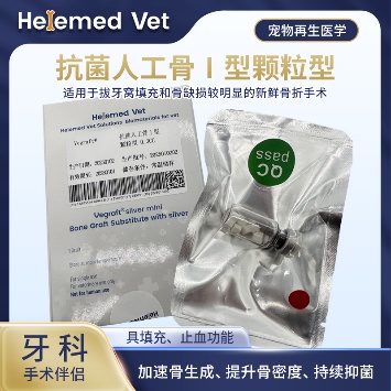
Antibacterial Artificial Bone Type I (Granules)
Alveolar bone filling material for dental applications, periodontal regeneration, alveolar bone reconstruction after extraction and bone-graft (ECM bone transplanted materials) procedures; induces autologous bone regeneration, works as a hemostatic material, inhibits infection and fills defects for a bone-graft effect.
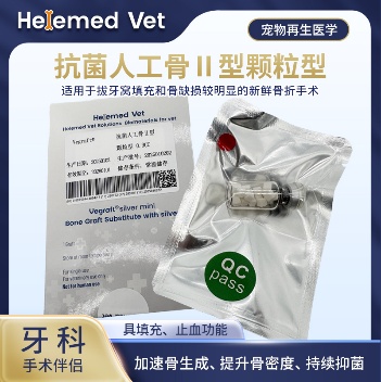
Antibacterial Artificial Bone Type II
Large-size dental ECM artificial bone material and alveolar bone filling material: periodontal regeneration, GBR guided bone regeneration and alveolar bone reconstruction after extraction; more economical for multiple teeth and repeated surgeries.
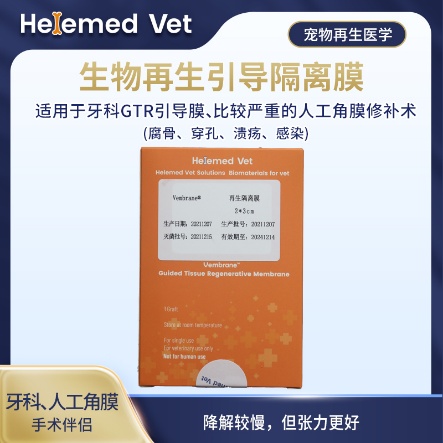
Bio-Guided Regeneration Membrane (2×3cm)
Double-layer GTR isolation membrane and GBR barrier membrane — a regenerative barrier membrane for dental guided tissue regeneration (GTR) and guided bone regeneration (GBR), plus more severe artificial corneal repair (sequestrum, perforation, ulcer, infection); slower degradation but better tension.
Supplied to veterinary clinics and hospitals. Ask your veterinarian whether periodontal regeneration or site preservation is appropriate for your dog.
8. Clinical cases
Three cases from our partner hospitals: the standard extraction-and-reconstruction workflow with the graft and the barrier membrane in place, guided tissue regeneration used to save the teeth of a ten-year-old dog whose owner declined multiple extractions, and alveolar bone reconstruction after extraction with radiographic follow-up.
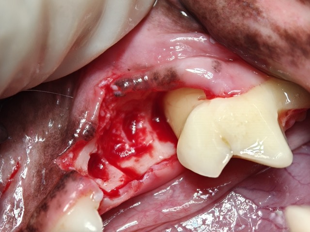
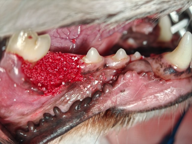
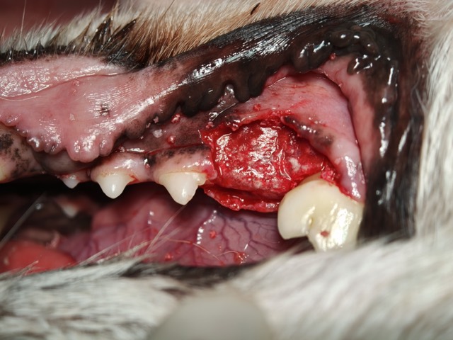
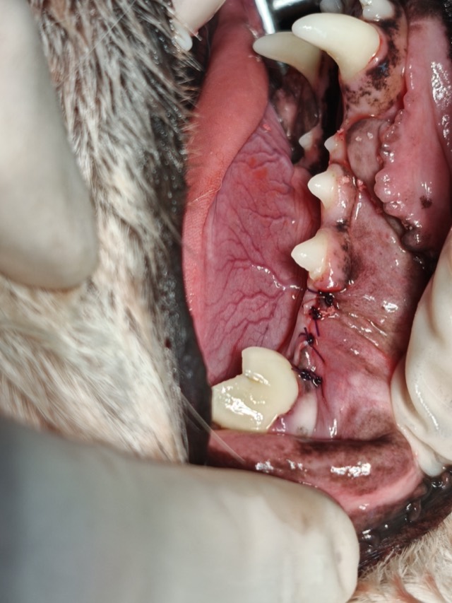


Standard Four Steps of Alveolar Bone Reconstruction
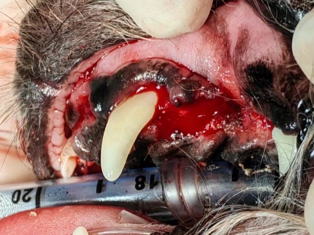
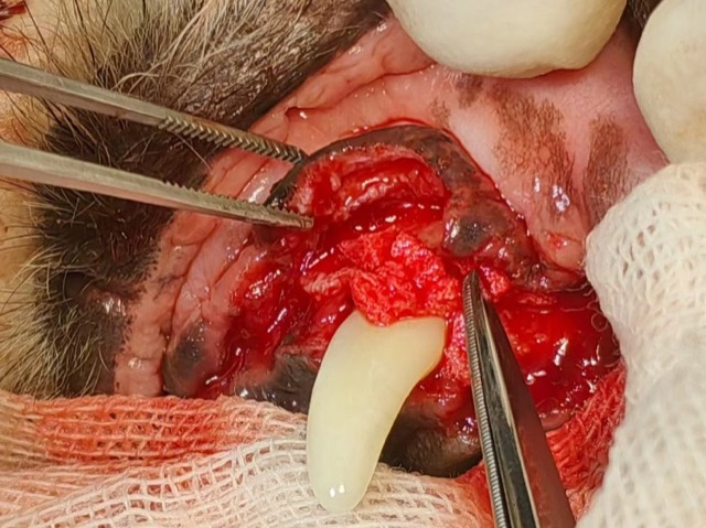
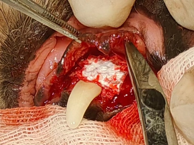
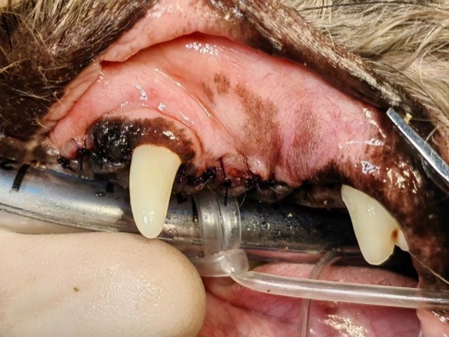
Standard workflow for extraction and alveolar bone reconstruction (site preservation surgery): ① extract and curette → ② fill Antibacterial Artificial Bone → ③ cover with Regeneration Barrier Membrane (isolating soft tissue) → ④ suture gingiva. Antibacterial Artificial Bone fills the socket, the membrane covers and isolates it from soft tissue, then gingiva is sutured — the same sequence used for subgingival regeneration and periodontal tissue regeneration; re-check at 3 months showed perfect, complete mandibular reconstruction.
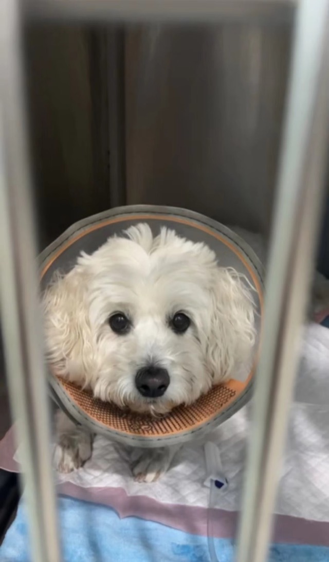
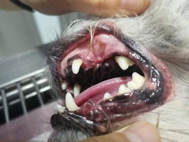
Periodontal GTR Guided Regeneration (Tooth-Saving)
A 10-year-old dog with severe periodontal disease — swollen and inflamed gums and alveolar bone atrophy; the owner declined multiple extractions and chose GTR tooth preservation (GTR tooth-saving surgery): mucosal flap reflection, extraction of the periapical-periodontitis tooth, root planing and repeated curettage, Antibacterial Artificial Bone granules packed and covered with Regeneration Barrier Membrane (smooth side inward), tension-free nodular suturing, and “button” principle to tighten the periodontal space around the canine. The goal of the procedure was periodontal regeneration, subgingival regeneration and periodontal tissue regeneration at the same site. At 10-day suture removal tooth stability was better, with good follow-up.
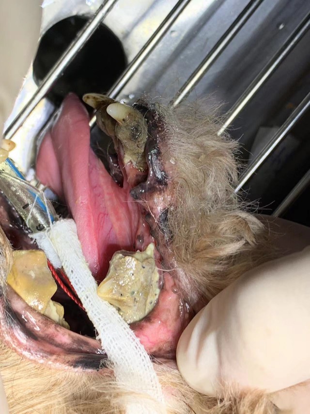
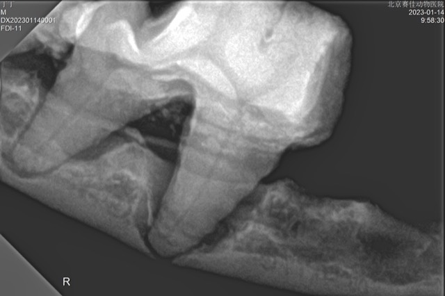
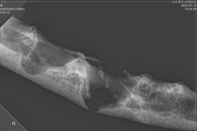
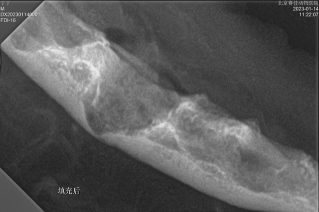
Tooth Extraction and Alveolar Bone Reconstruction
After extraction, site preservation surgery is needed: alveolar bone regeneration and alveolar bone reconstruction prevent fracture, oronasal fistula, facial fistula and other risks. X-ray showed mandibular dissolution and defect; after filling with Antibacterial Artificial Bone granules (alveolar bone filling material) the alveolar bone was regenerated. No infection or fracture post-op, and re-check showed good mandibular regeneration.
Cases from partner veterinary hospitals. ECM materials are veterinary surgical materials and are applied by the treating veterinarian. See all 39 clinical cases →
Frequently asked questions
Can dog periodontal disease be cured?
Gingivitis can be cured — at that stage the damage is reversible with a professional clean and daily home care. Once attachment and bone have been lost, the disease cannot be reversed; it can only be controlled. That means a professional clean at the interval your vet recommends, home care in between, and monitoring of the teeth that already have pockets.
How often does a dog need a dental cleaning?
It depends on the dog, the breed and how well home care is done. Many dogs need a professional clean and assessment once a year; small breeds with crowded mouths, and dogs whose owners cannot brush daily, often need one more frequently. The signs that the interval has run out are tartar returning at the gum line, bad breath coming back, and red gum margins.
Is anaesthesia-free dental cleaning safe or effective?
No, for two reasons. It removes only the tartar above the gum line and leaves the subgingival plaque and tartar that cause the disease, so the mouth looks better while the process continues. And scaling a conscious, moving patient carries a real risk of injury to the gum. A dental assessment that does not include radiographs and probing below the gum line is not an assessment of the disease.
Does brushing a dog’s teeth really make a difference?
It is the most effective thing an owner can do. Plaque is soft when it first forms and can be brushed away; once it mineralises into tartar it has to be scaled off. Daily brushing interrupts that cycle before it starts. Most dogs accept it if it is introduced gradually, and pet toothpaste comes in flavours designed for the purpose — human toothpaste must never be used, because fluoride and xylitol are toxic to dogs.
My dog has bad breath but eats normally — does that mean the teeth are fine?
No. Dogs hide dental pain, and appetites often stay normal with several painful teeth. Bad breath means bacteria and inflammation are already present at the gum line. The reliable way to know what is happening is an examination with probing and dental radiographs under anaesthesia, since the most damaged part of the tooth and the supporting bone are below the gum and cannot be seen from the outside.
When should a damaged tooth be saved rather than extracted?
Retention is reasonable when attachment loss is limited, the pockets can be cleaned, the dog can be rechecked, and the owner will maintain the tooth at home. Treatment then means lifting the gum, cleaning the root surface under direct vision, and in suitable cases placing a graft with a barrier membrane so that periodontal tissue and bone can regenerate — guided tissue regeneration. When loss is severe, the tooth is mobile, or the owner cannot maintain it, extraction is usually the kinder option.
Can dental disease affect the rest of my dog’s body?
Yes. A periodontal pocket is an open, bacteria-filled wound sitting on a rich blood supply, and chewing pushes bacteria into the bloodstream. Chronic oral infection is associated with changes in the kidneys and liver, with heart valve disease, and with poorer control of diabetes. Treating the mouth is not cosmetic work.