
- 1. Why a corneal ulcer is an emergency
- 2. What a corneal ulcer actually is
- 3. Signs, and how the vet confirms it
- 4. Depth decides everything
- 5. Medical treatment, and your part in it
- 6. What makes an ulcer worse
- 7. How long healing takes
- 8. When surgery is needed
- 9. Clinical cases
- Frequently asked questions
- Related guides
1. Why a corneal ulcer is an emergency
The cornea is the most exposed tissue in the body and the least able to defend itself:
- It has no blood vessels. The cornea is nourished by tears on the outside and by fluid from inside the eye. With no blood supply, it cannot mount the fast inflammatory response that skin or muscle can, and it cannot deliver immune cells to a wound quickly.
- Its own repair cells are slow. The corneal layer regrows from the edges of the defect, at roughly a millimetre a day at best. A large ulcer is therefore a race between the surface closing and the infection going deeper.
- Bacteria release collagenases. These enzymes digest the corneal tissue itself. This is what a melting ulcer is: the cornea being actively dissolved, sometimes visibly changing shape between one recheck and the next.
- Perforation is a one-way door. Once the cornea gives way, the fluid inside the eye leaks out, the eye collapses, and the choice narrows to emergency surgical repair or removal of the eye.
Seen today, not tomorrow: the eye is held closed or the dog squints constantly · the cornea looks cloudy, grey-blue or white · the dog paws at the face or rubs the eye on furniture · a visible dent, rough patch or dark spot on the eye surface · the eye looks larger than the other one · blood inside the eye. A painful eye is an emergency because the dog cannot tell you how much it hurts, and because the damage goes on while it waits.
One more reason not to wait: pain drives rubbing, and rubbing drives damage. A dog with an untreated ulcer will scratch at the eye with a hind foot and turn a surface defect into a deep one overnight.
2. What a corneal ulcer actually is
The cornea is built in layers, and the name of the ulcer is really a statement about how far down the damage goes:
- Epithelium — the outer skin of the eye, a few cell layers thick, and the layer that regrows fastest.
- Stroma — the thick middle layer, mostly collagen, and the layer that gives the cornea its strength and clarity. When stroma is lost, the eye both weakens and scars.
- Descemet’s membrane — a thin, very tough sheet that sits under the stroma. An ulcer that reaches it is called a descemetocele, and the eye is held together by that sheet alone.
- Endothelium — a single cell layer on the inside that pumps fluid out of the cornea to keep it clear. If it is damaged, the cornea stays cloudy permanently.
Most ulcers in dogs start as trauma: a cat scratch, a grass seed or foreign body under the eyelid, a chemical burn, or an eyelash that rubs. A smaller but important group starts with a condition that keeps the surface unhealthy — dry eye, an eyelid that rolls in (entropion), extra eyelashes (distichiasis), or a breed predisposition. Infection is usually secondary: the ulcer opens the door, the bacteria walk in.
That distinction is the reason treatment is not just "an antibiotic drop". If the underlying cause — dry eye, a rubbing eyelid, a grass seed — is not dealt with, the ulcer either does not heal or comes back.
3. Signs, and how the vet confirms it
What owners notice, roughly in order of how often it is the presenting complaint:
- Squinting and holding the eye closed, sometimes with the third eyelid partly raised over the eye
- Watering — the eye runs constantly, and the fur below it stays wet
- Pawing at the face or rubbing the eye along the floor or furniture
- A grey, blue or white patch on the eye, or the whole cornea looking hazy
- Reluctance to be handled around the head, or turning the head away from light
- A red, bloodshot eye, with the blood vessels around the cornea standing out
The examination that matters is the fluorescein stain. A drop of yellow dye is placed on the eye and the eye is rinsed; where the corneal surface is intact the dye washes off, and where tissue is missing the dye sticks to the exposed stroma. Under a cobalt blue light the defect shows up as a bright green area, and its size and shape can be drawn and compared at every recheck.
Alongside the stain, the vet will usually do:
- Schirmer tear test — a paper strip in the lower lid for 60 seconds, to find dry eye, which is one of the commonest reasons an ulcer will not heal
- Slit lamp or magnified examination — to judge the depth and look for a foreign body or a hair rubbing the surface
- Tonometry — to check the pressure inside the eye, since an ulcer and glaucoma can occur together
- Cytology, and culture if the ulcer is infected or melting — to identify the organism and stop guessing at antibiotics
The rule that follows from the stain: a steroid drop must never be used on an eye that has not been stained. Steroids switch off the repair response and increase collagenase activity, and on an ulcer that means the cornea starts to melt. Every case of a perforated eye caused by drops, rather than by the injury itself, traces back to this.
4. Depth decides everything
Two ulcers of the same size can need completely different treatment, because it is the depth that determines whether the eye can heal itself or has to be supported surgically:
| Ulcer | What has been lost | How it is managed |
|---|---|---|
| Superficial | Epithelium only; the stroma is intact and the eye is still strong | Antibiotic drops, pain relief, protection and a cone, with re-staining every few days. Most heal in seven to ten days. |
| Deep stromal | A significant part of the stroma; the eye looks hazy and is very painful | Intensive medical treatment around the clock, and surgical support is often needed to hold the surface while it heals. |
| Melting | Stroma being actively digested by collagenases from bacteria and from the cornea itself | A true emergency. Intensive treatment including serum and sometimes surgery; the cornea can perforate within hours. |
| Descemetocele | Everything except Descemet’s membrane; the eye is bulging under its own pressure | Surgery now. A graft, a flap or a biological patch is used to hold the eye together. |
| Perforation | The cornea has given way and fluid is leaking from inside the eye | Emergency surgical repair, or removal of the eye if it cannot be saved. |
An ulcer also changes depth in both directions. With good treatment a superficial ulcer closes and the stain becomes negative. Without it — or with the wrong drop, or with the dog rubbing the eye — a superficial ulcer becomes a deep one, then a melting one, often within 24 to 48 hours. That speed is the whole reason this is treated as urgent rather than as a wait-and-see problem.
5. Medical treatment, and your part in it
What the vet puts in place for an uncomplicated ulcer:
- A topical antibiotic to control secondary infection. The choice depends on whether the ulcer is infected, what grew on culture, and how often you can realistically medicate the eye.
- Pain relief. Topical atropine relieves the painful spasm of the pupil, and a systemic anti-inflammatory or other painkiller controls the rest. Pain relief is not a comfort measure here; it is part of the treatment, because a comfortable dog stops rubbing the eye.
- Serum drops, in melting or infected ulcers. Serum from the dog’s own blood contains antiproteinases that work against the enzymes digesting the cornea, and it is prepared fresh by the clinic.
- Protection of the surface — a bandage contact lens, a temporary tarsorrhaphy where the eyelids are partly sutured shut, or in some cases a conjunctival flap. The point is to give the surface a protected, moist environment and stop the eyelid from scraping across the defect.
- An Elizabethan collar, worn continuously. Not optional, and not only while you are watching. Most ulcer complications happen overnight or when the dog is alone.
- Re-staining at intervals so that improvement is measured and not assumed.
What you are responsible for at home is narrower than owners expect, and more demanding:
- Give every dose on time. Ulcers are often treated every two to six hours at the start. A missed dose in a melting ulcer matters; a missed day can undo the treatment.
- Observe any storage instruction. Some drops and serum must be refrigerated, and some have a short life once opened.
- Keep the collar on and check that it is not so loose the dog can reach the eye around it.
- Do not bathe the dog, and keep shampoo and water out of the eye while the ulcer is open.
- Return for the recheck even if the eye looks better. An eye that looks comfortable can still have a staining defect, and the treatment is stepped down on the basis of the stain, not of appearance.
6. What makes an ulcer worse
Almost every ulcer that turns into a lost eye does so because of something on this list:
- A steroid eye drop used before a fluorescein stain. This is the single most destructive thing an owner or a well-meaning clinic can do. The cornea melts, and the ulcer that was treatable becomes surgical.
- Leftover human eye drops, or drops prescribed for another dog or for yourself. The concentration, the preservative and the drug itself may all be wrong for a dog with an open ulcer.
- Human skin ointment or antibiotic cream applied to the eye. The base is not made for the cornea and can cause a chemical injury on top of the ulcer.
- Rubbing. A dog left without a collar, or with inadequate pain relief, will physically re-injure the eye. This is the commonest reason an ulcer goes from superficial to deep in one night.
- Home-made salt solutions or repeated flushing with tap water, which are neither sterile nor the right salt concentration and irritate the surface further.
- Removing a foreign body yourself, or trying to push a protruding mass back into place. Flushing gently with sterile saline while you travel to the clinic is the limit of what to attempt.
- Waiting to see if it improves. The size of the defect can be measured at every appointment; the cost of waiting is not a week of discomfort, it is the difference between drops and surgery.
If a steroid drop has already been given, do not wait to see what happens. Take the bottle with you, tell the vet exactly what was applied and when, and have the eye stained immediately. That information changes the treatment plan on the spot.
7. How long healing takes
Owners want a single number, and the honest answer is a range that depends on depth, cause and cooperation:
| Situation | Typical healing time | What you will see |
|---|---|---|
| Superficial ulcer, no complications | 7–10 days | The stain area shrinks at each recheck, then goes negative. Treatment usually continues for about a week after the stain is clean. |
| Superficial ulcer with dry eye or a lid problem | Weeks, and it may recur | Slow or stalled healing until the underlying cause is treated. The eye may look stable and then break down again. |
| Deep stromal ulcer | Weeks | A slower closure, more scarring, and often several rechecks with the stain. |
| Melting ulcer | Days to weeks, with surgery | Rapid change in the first 48 hours, then stabilisation. The priority is to stop the melting, not to close the defect. |
| Surgically repaired cornea | Weeks to months | Healing under a graft or patch, with the surface closing beneath it and the material degrading as the corneal layer regenerates. |
Two things owners find alarming and should not: blood vessels growing into the cornea, which look like red threads reaching across the eye, are a good sign — they mean the eye has managed to bring a blood supply to the repairing area, and they usually fade once healing is complete. And a white or grey haze left behind after the ulcer closes is scar tissue in the stroma; it often becomes less obvious over months, though a deep ulcer can leave a permanent opacity.
The reasons healing stalls are worth knowing, because each has an answer: dry eye that is not being treated, a foreign body or an eyelash still rubbing the surface, an infection that is not controlled by the antibiotic chosen, an eyelid that rolls in, a dog that is still getting at the eye, and a cornea that has been treated with a steroid. A dog whose ulcer is not shrinking at the seven-day recheck needs to be re-examined for these rather than simply continued on the same drops.
8. When surgery is needed
Surgery is considered when the eye cannot close the defect on its own — a deep or melting ulcer, a descemetocele, a perforation, or an ulcer that has failed to heal after correct medical treatment. The options, in the order they are usually considered:
- A conjunctival flap or graft — a piece of the dog’s own conjunctiva is rotated over the defect and sutured in place. It brings a blood supply directly to the area and is robust and well established; the trade-off is that it is opaque, so the eye is covered while it heals.
- A corneal graft — donor cornea is sutured into the defect to restore the surface, and is used where the aim is to preserve clarity as well as strength.
- A biological scaffold patch — a thin, absorbable sheet, such as an ECM membrane, is placed over the defect as a barrier and as a scaffold that the corneal layer grows into. Because it is thin, and because it degrades as the cornea regenerates rather than staying in place, it is used where the conjunctival flap would be unnecessarily bulky, and in eyes where some clarity is worth preserving.
- Amniotic membrane or a combination technique, where the defect has to be filled and then covered.
Three practical points decide whether the repair works:
- The defect must be clean and free of infected or necrotic tissue before anything is placed — no patch tolerates a contaminated bed.
- Orientation matters. For a scaffold patch, the rough side faces the eyeball and the smooth side faces outward, and the surgeon sets this deliberately. It is not interchangeable.
- The patch protects, it does not replace treatment. Antibiotic drops, pain relief and the collar continue after surgery, and the eye is re-examined at intervals during healing.
And one point about what surgery is not: it is not a way to avoid the diagnosis. The best outcome for a cornea that has lost tissue is achieved at the first attempt, on an eye that was stained, assessed and treated correctly from the start.
Materials used in corneal repair
Our ECM regenerative materials are veterinary surgical materials, applied by the treating veterinarian after the eye has been examined and stained. The Bio-Guided Regeneration Membrane is used as a corneal patch: the 20×20mm single layer is the thinnest sheet in the series and is used for milder defects, while the 20×30mm double layer degrades more slowly and has greater tensile strength, and is used for more severe repair such as perforation, corneal sequestrum, deep ulcer and established infection. ECM Antibacterial Powder has also been applied to the corneal layer under a bandage contact lens, with the eyelids partially sutured for protection.
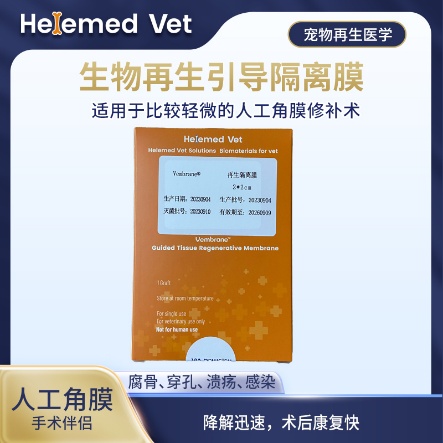
Bio-Guided Regeneration Membrane (2×2cm)
Corneal patch: for milder artificial corneal repair (sequestrum, perforation, ulcer, infection); fast degradation and quick post-op recovery, so it is also the thinnest regenerative isolation membrane in the series.
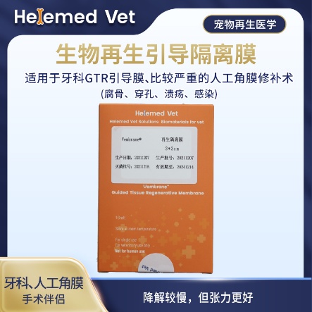
Bio-Guided Regeneration Membrane (2×3cm)
Double-layer GTR isolation membrane and GBR barrier membrane — a regenerative barrier membrane for dental guided tissue regeneration (GTR) and guided bone regeneration (GBR), plus more severe artificial corneal repair (sequestrum, perforation, ulcer, infection); slower degradation but better tension.
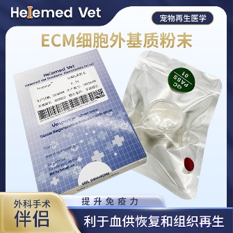
ECM Antibacterial Powder
ECM bioscaffold powder — an extracellular matrix (ECM) based antibacterial and inhibitory powder: widely used as ECM trauma dressings on infected internal/external wounds, fresh wounds, burns and crush injuries to markedly improve blood supply, accelerate healing, inhibit infection and reduce complications; it is also an ECM hemostatic material that stops capillary bleeding. How to use: after debridement, take a small amount of powder with forceps and apply to the wound surface, moisten with saline (moist wound healing) and bandage; apply once every 5 days — daily debridement is not required.
Supplied to veterinary clinics and hospitals. These are surgical materials used by the treating veterinarian as part of a repair procedure — they are not eye drops and cannot be used at home.
9. Clinical cases
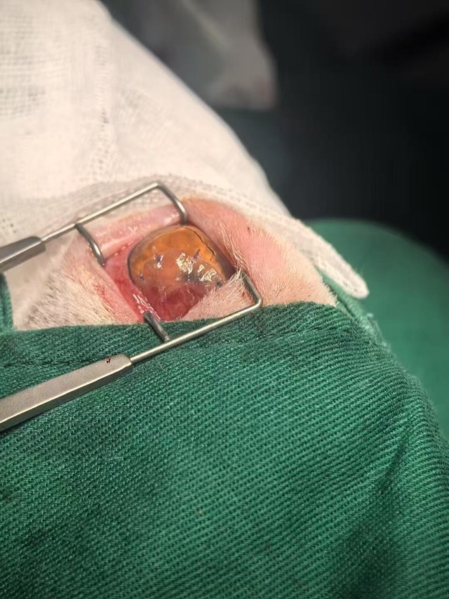
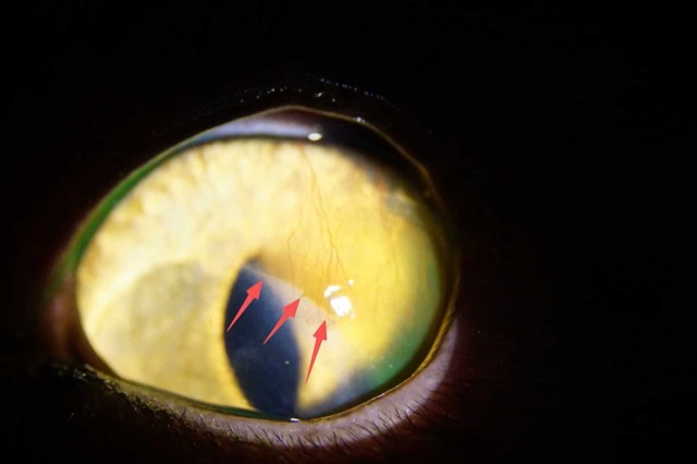
Three eyes from our partner hospitals, all of which had already lost corneal tissue by the time they were seen: an ECM membrane used to rebuild the corneal layer, a surgical repair where the patch’s rough side faced the eyeball and the smooth side outward, and an antibacterial powder application to the cornea protected under a contact lens and eyelid sutures.
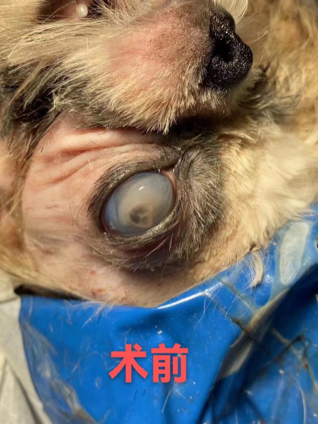
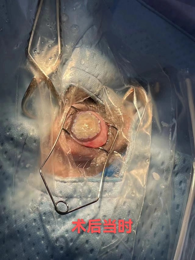
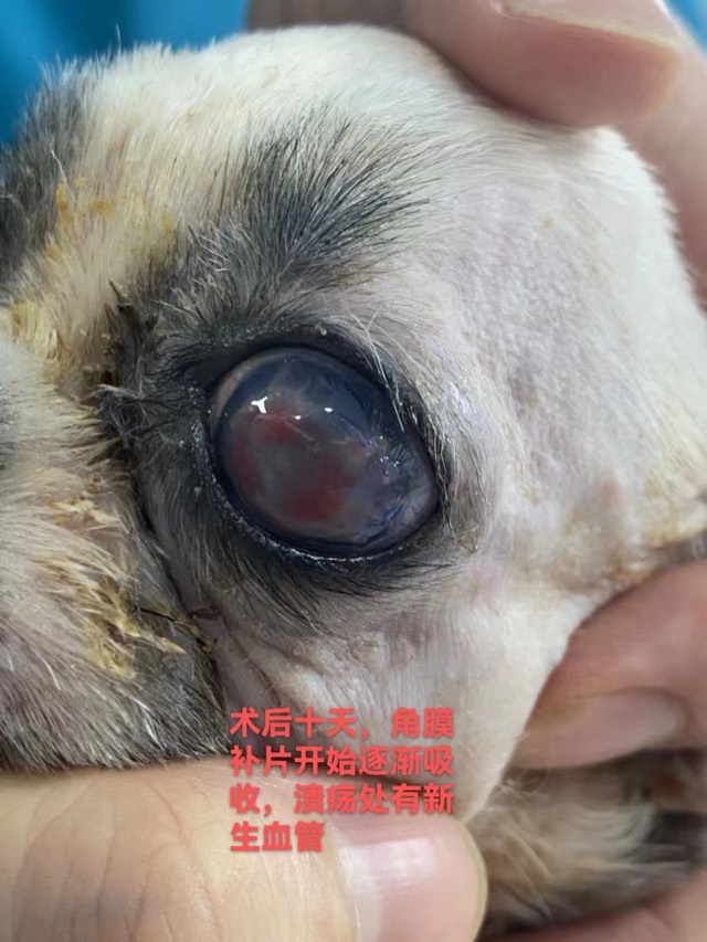

Corneal Patch Transplantation — Corneal Recovery
The ECM membrane sheet (Corneal Patch) is used for corneal-layer repair such as ulcers and perforations: as an inductive scaffold for the corneal layer, recruiting cells and vessels to grow in and inducing corneal regeneration.
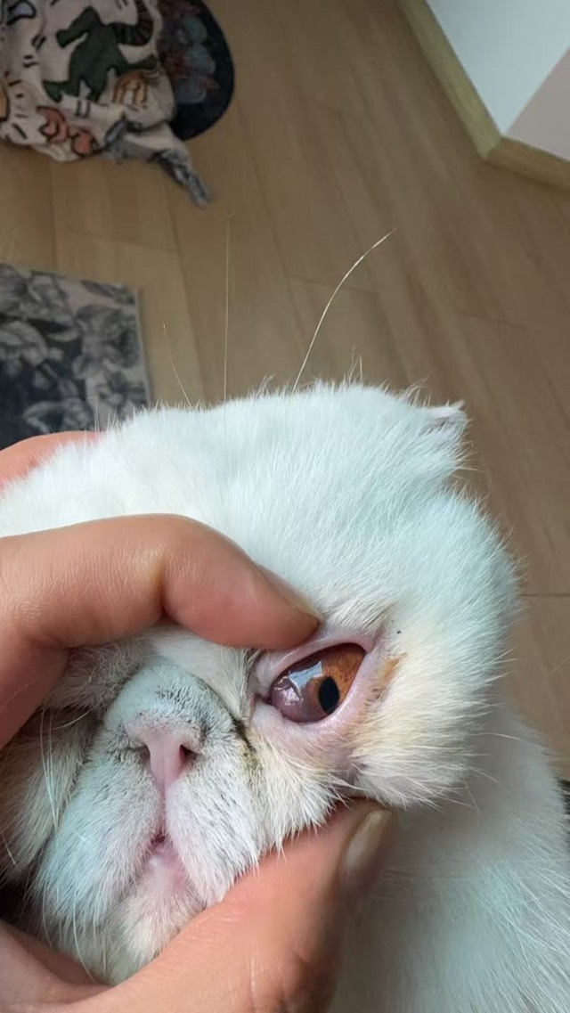
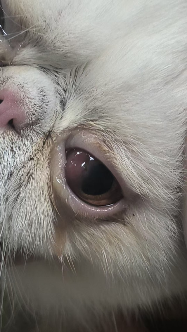
Corneal Patch Transplantation — Surgical Repair
Corneal repair surgery on Apr 27, 2025: the Corneal Patch's rough side faced the eyeball and smooth side outward; feedback on May 15 said corneal growth was very good, better than conjunctival-flap coverage.

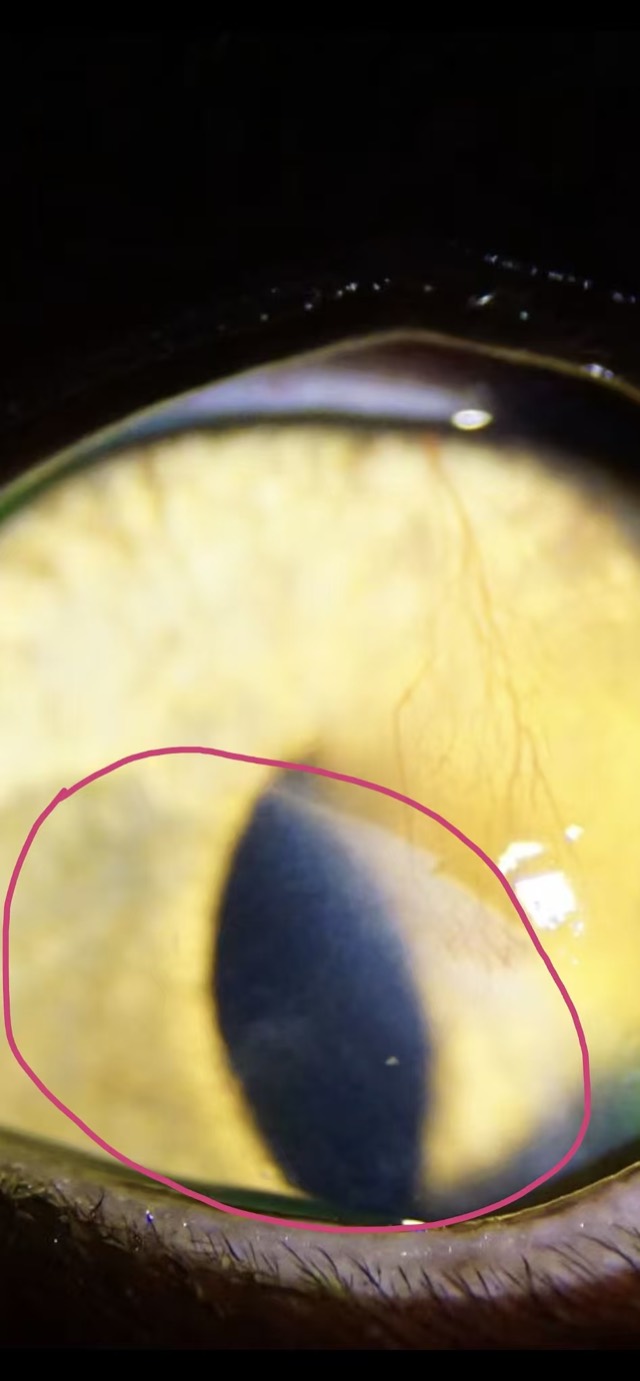
ECM Antibacterial Powder Corneal Application
ECM Antibacterial Powder was applied on the corneal layer with a bandage contact lens and eyelid suturing for protection; the covered area fully healed with a clear boundary line from the untreated area (outside the red circle is the untreated area).
Cases from partner veterinary hospitals. ECM materials are veterinary surgical materials and are applied by the treating veterinarian. See all 39 clinical cases →
Frequently asked questions
How long does a dog corneal ulcer take to heal?
A superficial ulcer with no underlying problem usually heals in seven to ten days, and treatment continues for about a week after the fluorescein stain becomes negative. Deep or melting ulcers take weeks, and an ulcer in an eye with untreated dry eye or a rubbing eyelid can stall for as long as the cause is left alone. Healing is judged by the stain shrinking at each recheck, not by how the eye looks.
Can I use human eye drops on a dog with an ulcer?
No, and one group is genuinely dangerous. A steroid drop, or a combined antibiotic-and-steroid drop, given on an ulcer accelerates collagen breakdown and can cause the cornea to melt and perforate. Never use drops from your own medicine cabinet, drops prescribed for another animal, or human skin ointment on a dog’s eye — and never use a steroid drop before a vet has stained the eye.
What is the fluorescein stain for?
It is a yellow dye that sticks only to corneal tissue where the surface has been lost, showing up bright green under a blue light. It is how an ulcer is confirmed rather than guessed at, how its exact size is recorded so progress can be measured at each recheck, and the test that must be done before a steroid is even considered.
Will my dog’s vision be affected after a corneal ulcer heals?
Often not much. A superficial ulcer usually heals with little or no lasting mark. A deep ulcer can leave a white or grey haze where scar tissue formed in the stroma, which may reduce clarity, though it frequently becomes less obvious over months. Blood vessels growing across the cornea during healing look dramatic but are a normal part of repair and usually fade once healing is complete.
Why does my dog have to wear a cone for an eye ulcer?
Because rubbing is what turns a surface ulcer into a deep one, and it usually happens when nobody is watching. Pain relief reduces the urge, but it does not remove it. The collar is part of the treatment, not a precaution, and it should stay on continuously until the vet confirms the ulcer has closed.
When does a corneal ulcer need surgery?
When the eye cannot close the defect on its own: a deep stromal ulcer, a melting ulcer, a descemetocele where only Descemet’s membrane is left, a perforation, or an ulcer that has failed to heal after correct medical treatment. Options include a conjunctival flap, a corneal graft or a thin absorbable biological patch, and the choice depends on how much tissue has been lost and whether clarity is worth preserving.
Can a dog get a corneal ulcer again?
Yes, if the underlying cause is still there. Dry eye, an eyelid that rolls in, extra eyelashes, a breed with shallow eye sockets and an exposed corneal surface all make repeat ulcers likely. That is why the recheck after healing matters: the point is not only to confirm the eye has closed, but to decide whether the dog needs ongoing lubrication, an eyelid correction or another long-term measure.