
- 1. What a dog ear infection actually is
- 2. Signs: what you see, smell and feel
- 3. Bacteria, yeast, mites or something else
- 4. Why it happens, and the allergy link
- 5. Why some dogs are built for ear trouble
- 6. How a vet examines the ear
- 7. Cleaning, drops and finishing the course
- 8. Why it keeps coming back
- 9. When an ear infection needs surgery
- 10. When the ear skin breaks down
- 11. Clinical cases
- Frequently asked questions
- Related guides
1. What a dog ear infection actually is
The medical term is otitis externa: inflammation of the external ear canal, the part from the opening to the eardrum. In dogs it is overwhelmingly a mixed problem — a surface that has become inflamed, and organisms that have multiplied in it. The canal becomes swollen, it produces more wax and discharge, and the discharge makes the skin more inflamed. That loop is why an ear that is "just a bit dirty" becomes an ear the dog cannot stop shaking.
Vets divide the causes into three layers, and it is the most useful way to think about the problem:
- Primary causes start the disease: allergy (atopy and food allergy), ear mites, a foreign body such as a grass awn, hormonal disease, or a disorder of the skin itself.
- Predisposing factors make it easier to start: a pendulous ear that traps moisture, hair growing inside the canal, narrow or hairy canals, frequent swimming, and the shape of the ear in certain breeds.
- Perpetuating factors keep it going once it has started: bacterial and yeast infection, a middle-ear infection behind an intact or ruptured eardrum, and irreversible thickening and narrowing of the canal.
The practical consequence is simple. An ear infection that only gets drops addresses the third layer and leaves the first two untouched — which is exactly the pattern of a dog that is treated four times a year for the same ear.
2. Signs: what you see, smell and feel
- Head shaking and repeated scratching at one or both ears — often the very first sign
- A smell you notice from across the room. A musty, yeasty odour points towards Malassezia; a sharper, pus-like smell points towards bacteria
- Discharge: dark brown or black crumbly debris, yellow pus, or a wet, waxy coating on the inside of the ear flap
- Redness and swelling of the ear flap and canal, sometimes with crusting at the opening
- Pain. The dog pulls away, tilts the head away from the ear, or yelps when the ear is touched. A dog that will not let you near the ear for cleaning should be seen by a vet before you try again
- Head tilt or a drooping ear flap, which suggest the ear is painful or the middle ear is involved
- A swollen, squishy ear flap — that is an ear hematoma, a pocket of blood between the cartilage layers, caused by violent head shaking
- Hearing changes, which owners rarely notice early: the dog stops responding to quiet sounds
One-sided signs are usually a local problem — a foreign body, a tumour, a polyp, or a single ear infection. Signs in both ears, or a history of three or more episodes, should send the vet looking for an allergy or a hormonal disease rather than another bottle of drops.
3. Bacteria, yeast, mites or something else
| What it is | Typical picture | How it is confirmed |
|---|---|---|
| Bacterial otitis — usually Staphylococcus pseudintermedius, in chronic cases often Pseudomonas aeruginosa | Red, painful canal; yellow to brown pus; strong smell; often worsens quickly | Cytology (stained smear), and culture for chronic or resistant cases |
| Yeast otitis — Malassezia pachydermatis | Greasy, thickened, dark canal; thick brown-black debris; musty smell; intense itch | Cytology — yeast cells are easy to see under the microscope |
| Ear mites — Otodectes cynotis | Very itchy, "coffee grounds" debris, usually in young dogs and in cats in the household | Otoscopy, or seeing the mites on a swab |
| Foreign body (grass awn, seed, hair) | Sudden, violent onset in one ear, often after a walk or in the summer | Otoscopy — the only way to see past the bend |
| Polyp or tumour | Chronic one-sided discharge, a visible mass, or a canal that will not clear | Otoscopy, biopsy |
| Middle-ear infection (otitis media) | Head tilt, pain when the mouth is opened, a ruptured eardrum, or an ear that flares again days after treatment stops | Otoscopy, imaging |
In practice the most common finding is both bacteria and yeast in the same ear. Treating only one of them is one of the reasons an ear improves for a week and then relapses.
4. Why it happens, and the allergy link
In puppy ears, the usual culprit is ear mites or a first infection. In adult dogs with recurrent ear disease, the usual culprit is allergy — and it is worth being blunt about it, because this is the single most missed step in ear treatment.
- Environmental allergy (atopy) is the most common underlying cause of recurrent ear disease in dogs. The ear canal is skin, and it reacts to the same allergens as the rest of the body. Many allergic dogs show ear signs first and skin signs later.
- Food allergy commonly affects ears — often both, often with gut signs and itch around the face, paws and rear.
- Flea allergy and other skin disease can also drive it.
- Hormonal disease — hypothyroidism and Cushing's disease — predisposes to skin and ear infection.
- Anatomy and lifestyle — swimming, humid weather, and a coat that traps moisture.
This is why an ear that has been treated more than twice in a year deserves a proper work-up rather than a third course of drops. The question stops being "which ear medication" and becomes "why is this ear repeatedly infected".
5. Why some dogs are built for ear trouble
Some breeds are over-represented in ear disease, and the reasons are structural rather than bad luck:
- Pendulous ears that cover the canal and reduce airflow — Cocker Spaniels, Basset Hounds, Beagles, Springer Spaniels.
- Hairy ear canals that hold wax and moisture — Poodles, Schnauzers, Shih Tzus, and many terriers.
- Narrow or stenotic canals — Shar-Peis and Chow Chows have famously tight canals; chronic inflammation narrows a canal further.
- Skin folds and narrow ear openings — Bulldogs, Pugs, French Bulldogs.
- Allergic breed predisposition — many of the above, plus Labrador and Golden Retrievers, German Shepherds and West Highland White Terriers.
None of this means the dog is doomed. It means the ear has to be checked regularly, kept dry, and any flare treated early rather than late.
6. How a vet examines the ear
- A proper look, both ears. The vet compares them, checks the ear flap for hematoma or thickening, and looks at the ear canal opening for discharge and crusting.
- Otoscopy. A scope goes into the canal to see the vertical and horizontal parts, whether the eardrum is intact, whether there is a foreign body, polyp or mass, and how much the canal has narrowed. In a painful ear this may need sedation, and clipping the hair around the opening is often necessary first.
- Cytology — the single most useful test. A swab or impression is stained and examined under the microscope. This is what separates bacterial from yeast infection and tells the vet which treatment will actually work.
- Culture and sensitivity for chronic, recurrent or previously treated ears, where resistant organisms such as Pseudomonas are likely.
- Imaging when the middle ear is suspected, or where the canal cannot be examined properly.
- An allergy or hormonal work-up when the history fits — this is what changes the long-term outcome.
Key point: "just give me the usual ear drops" is how ears become resistant. Cytology takes minutes and it decides whether the problem is bacteria, yeast, both, or neither.
7. Cleaning, drops and finishing the course
- Clean the canal properly first. A medicated ear cleaner prescribed or supplied by your vet breaks up wax and debris so the medication can reach the skin. The ear is filled, the base of the ear is massaged, the dog shakes, and the loosened debris is wiped away from the opening with cotton wool. Cleaning is not optional; medication applied on top of a plug of wax does nothing.
- Apply the medicated drops for the full course. Most ear preparations combine an antibacterial, an antifungal and often a corticosteroid to bring down the swelling that is trapping everything in. Courses typically run two to four weeks — and longer where the canal is thickened or infection has been present for months. Many relapses happen because treatment stopped when the ear looked clean rather than when the infection had cleared.
- Recheck. A vet re-examines and re-does the cytology at the end of treatment. An ear that looks better is not the same as an ear that is clear.
- Treat the underlying cause. All-year flea control, an allergy plan, a proper diet trial, treatment of hormonal disease, and — where anatomy is the issue — a plan for managing a hairy or pendulous ear long-term.
- Control the pain and itch so the dog stops shaking the head and scratching. Violent head shaking is what causes an ear hematoma, and scratching keeps the canal inflamed.
What not to do: do not push cotton buds into the canal — you will pack debris further down and risk the eardrum. Do not use alcohol, hydrogen peroxide, vinegar mixtures or human ear preparations on a dog. Do not stop the drops early because the smell has gone. Do not use leftover antibiotic or steroid drops from a previous episode: the organism, or the mix of organisms, has usually changed.
8. Why it keeps coming back
- The course was too short for the degree of change in the canal. Chronically inflamed skin takes weeks to return to normal.
- The underlying allergy was never identified or managed, so the canal re-inflames as soon as treatment stops.
- A resistant organism was never cultured. Pseudomonas in particular is common in ears treated repeatedly and is not sensitive to the usual first-line drops.
- Middle-ear infection was missed. If the infection sits behind the eardrum, topical drops cannot reach it, and the ear relapses days after the drops stop.
- Perpetuating anatomical change was not addressed. Repeated inflammation thickens and narrows the canal and narrows the drainage — the ear can no longer clean itself.
- A foreign body or mass was never found, because the canal was too painful or too narrow to examine fully without sedation.
9. When an ear infection needs surgery
Most ear infections never need an operation. It becomes a surgical question in a defined set of situations, and it is worth knowing them, because owners often meet the word "surgery" late and assume it came out of nowhere:
- Ear hematoma (aural hematoma). Violent head shaking from an itchy or painful ear ruptures a blood vessel between the two layers of cartilage in the ear flap. Blood collects and the flap fills like a pillow. This is treated surgically — the pocket is opened, the clot and fibrin removed, and the two surfaces are apposed so they heal together — and the ear infection that caused the shaking has to be treated at the same time.
- Ear canal polyps and tumours, which block drainage and are removed.
- Chronic proliferative otitis externa — an ear canal whose skin has permanently thickened and narrowed from months or years of inflammation, closing off drainage. Options range from removing the diseased tissue while preserving hearing, through to total ear canal ablation where the canal is beyond salvage.
- Ear canal stenosis or atresia after chronic inflammation, trauma or previous surgery, where the canal must be reconstructed or removed.
- Middle-ear disease that does not respond to medical treatment, requiring the bony chamber to be opened and drained.
Ear surgery leaves a real wound: a cavity to be managed, a flap that must stay in place, and a site that is difficult to bandage. It is exactly the kind of wound that needs the infection controlled, dead space dealt with, and the surface covered while it heals.
10. When the ear skin breaks down
Chronic ear disease frequently damages the skin of the ear flap and the surrounding area — not just the canal. The mechanisms are the ones you would expect: constant scratching and rubbing, self-trauma during head shaking, maceration of skin from discharge, and surgery itself. The results are ulcers, open raw areas, and after surgery a cavity that has to be closed in from the inside out.
Once skin is missing rather than just inflamed, the priorities change from "which drops" to wound management:
- Control the infection, and get the discharge off the skin so it stops macerating more of it.
- Stop the self-trauma — collar, pain relief, and treating the itch.
- Deal with dead space. A cavity left after removal of a hematoma, a mass or a diseased canal will fill with fluid and re-open unless it is managed.
- Cover the defect so the surface can heal and remaining skin can migrate across it.
Materials used in ear and head wounds
Our ECM regenerative materials are veterinary surgical materials, applied by the treating veterinarian after the wound or cavity has been cleaned and debrided. ECM Sponge is used to fill and support a surgical cavity — the situation that follows hematoma surgery or removal of a diseased ear canal — while the antibacterial powder is applied to the debrided wound bed and dressed.
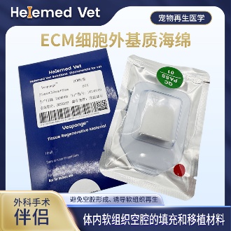
ECM Sponge
ECM filling sponge and ECM cavity-filling biomaterials: for internal soft-tissue cavity filling, providing long-lasting space occupation and inducing regeneration to achieve a soft-tissue graft effect; also an ideal ECM Tumor Filling Material for dead-space repair after tumor resection, and a routine companion to ECM trauma dressings in cavity and fistula wounds.
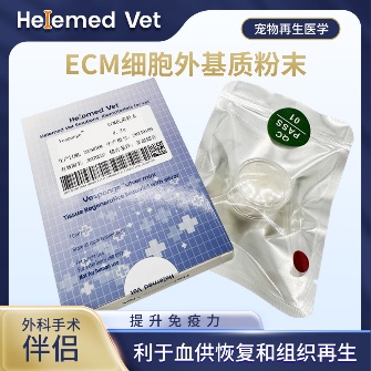
ECM Antibacterial Powder
ECM bioscaffold powder — an extracellular matrix (ECM) based antibacterial and inhibitory powder: widely used as ECM trauma dressings on infected internal/external wounds, fresh wounds, burns and crush injuries to markedly improve blood supply, accelerate healing, inhibit infection and reduce complications; it is also an ECM hemostatic material that stops capillary bleeding. How to use: after debridement, take a small amount of powder with forceps and apply to the wound surface, moisten with saline (moist wound healing) and bandage; apply once every 5 days — daily debridement is not required.
Ask your veterinarian whether these materials suit your dog's surgical site; they are supplied to veterinary clinics and hospitals.
11. Clinical cases
These two cases from our partner hospitals show ECM Sponge being used to fill a surgical cavity after tissue has been removed — the same problem a haematoma operation or an ear canal removal creates, where the space left behind has to be supported while it heals.
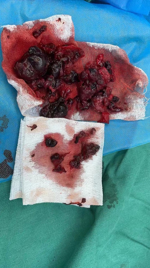
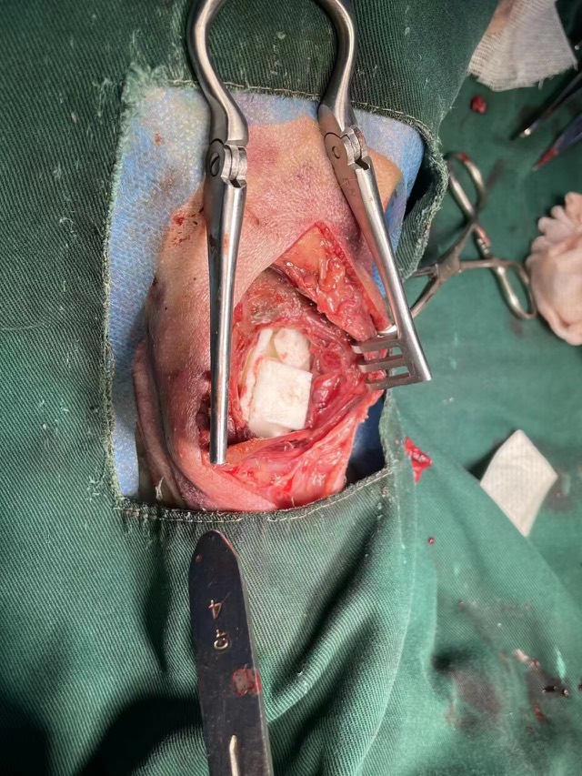
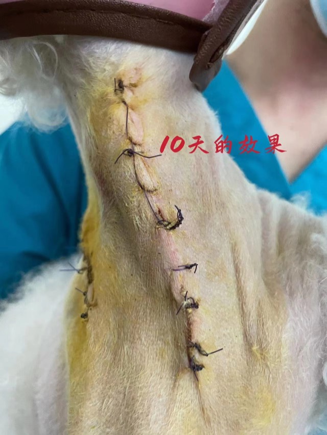
ECM Sponge Assisted Cavity Filling
When there is an obvious cavity with normal surrounding blood supply, ECM Sponge can assist filling (larger volume, ~20-day degradation, synchronized with new tissue). Before use, fully soak in saline and squeeze out excess water.
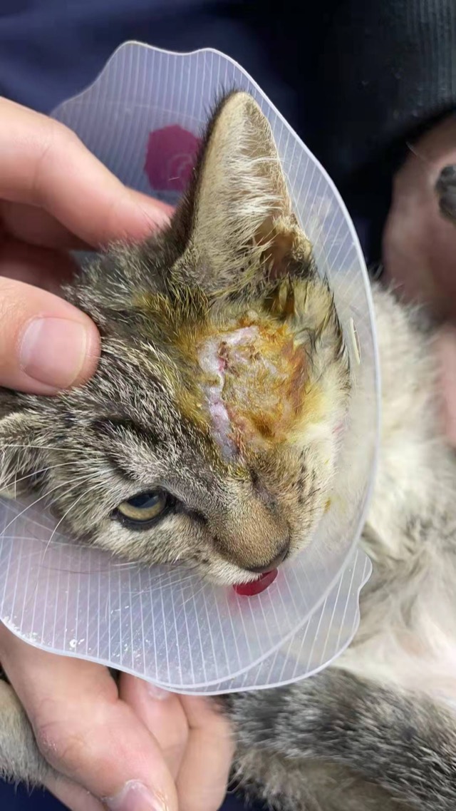
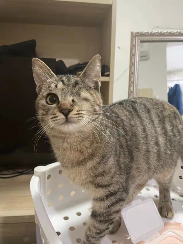
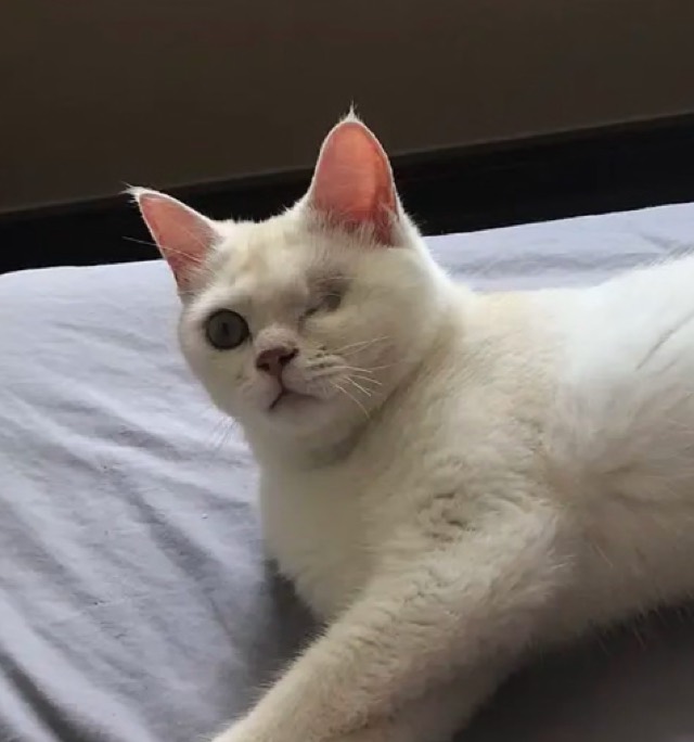
Enucleation Cavity Filling
After enucleation, ECM powder and ECM Sponge were placed to reduce exudate and speed recovery; at 2 months it remained full — the sponge had been replaced by autologous new tissue. Without filling there would inevitably be a depression and a permanent subcutaneous cavity. Apply moderately; more is not better.
Cases from partner veterinary hospitals. ECM materials are veterinary surgical materials and are applied by the treating veterinarian, not at home. See all 39 clinical cases →
Frequently asked questions
How do I know if my dog has an ear infection?
The early signs are head shaking, scratching at one ear, and a smell you notice without putting your nose to the ear. You may see brown, black or yellow discharge, redness, and pain when the ear is touched. A swollen, squishy ear flap is a hematoma and needs a vet.
Will a dog ear infection clear up on its own?
A mild, early flare can sometimes settle, but most ear infections in dogs become deeper and more painful if untreated, and the canal can narrow permanently. Because the underlying cause is usually an allergy, an untreated ear commonly returns. Have it examined rather than waiting it out.
Can I treat my dog’s ear infection at home?
You can and should clean the ear with a cleaner your vet has approved, and give the prescribed drops for the full course. You should not use hydrogen peroxide, alcohol, vinegar, olive oil or human ear drops, and you should not push cotton buds into the canal. Those measures either damage the skin or push debris towards the eardrum.
Why does my dog keep getting ear infections?
In adult dogs the usual reason is allergy — environmental or food — with the ear flare being the visible symptom. Other reasons include a course of drops that was too short, a resistant organism such as Pseudomonas, a missed middle-ear infection, or a canal that has permanently narrowed. Repeated ear infections need a work-up, not another bottle of the same drops.
Is a dog ear infection contagious to other pets or to people?
The bacterial and yeast infections found in a dog ear are not contagious in the usual sense. Ear mites are contagious between pets in the same household, so if mites are found, every pet should be treated.
When does an ear infection need surgery?
When the ear flap fills with blood (an aural hematoma), when a polyp or tumour blocks the canal, when chronic inflammation has permanently narrowed or closed the canal, or when a middle-ear infection does not respond to medical treatment. Most ear infections never reach this point.
How long does it take to treat a dog ear infection?
A simple infection usually needs two to four weeks of cleaning and medicated drops, with a recheck and repeat cytology at the end. Ears with thickened, narrowed canals, or with a middle-ear component, take considerably longer. Stopping when the ear looks clean is the most common reason for a relapse.